 Movie
Movie Controller

[English] 日本語
 Yorodumi
- PDB-5zo2: Crystal structure of mouse nectin-like molecule 4 (mNecl-4) full ... -
+ Open data

- Basic information
| Entry | Database: PDB / ID: 5zo2 | |||||||||
|---|---|---|---|---|---|---|---|---|---|---|
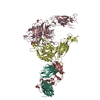
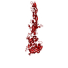
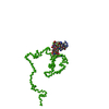
| Title | Crystal structure of mouse nectin-like molecule 4 (mNecl-4) full ectodomain in complex with mouse nectin-like molecule 1 (mNecl-1) Ig1 domain, 3.3A | |||||||||
 Components Components |
| |||||||||
 Keywords Keywords | CELL ADHESION / cell adhesion molecule / glycoprotein / Ig domain / SynCam / CADM / Nectin-like / Necl-4 / Necl-1 / axon / Schwann Cell / Molecular Replacement / myelogenesis / heterogeneous dimer | |||||||||
| Function / homology |  Function and homology information Function and homology informationnegative regulation of peptidyl-tyrosine phosphorylation / Nectin/Necl trans heterodimerization / negative regulation of vascular endothelial growth factor signaling pathway / vascular endothelial growth factor receptor 1 binding / Adherens junctions interactions / synaptic membrane adhesion / regulation of Rac protein signal transduction / negative regulation of peptidyl-threonine phosphorylation / cell-cell contact zone / regulation of wound healing ...negative regulation of peptidyl-tyrosine phosphorylation / Nectin/Necl trans heterodimerization / negative regulation of vascular endothelial growth factor signaling pathway / vascular endothelial growth factor receptor 1 binding / Adherens junctions interactions / synaptic membrane adhesion / regulation of Rac protein signal transduction / negative regulation of peptidyl-threonine phosphorylation / cell-cell contact zone / regulation of wound healing / regulation of cell motility / vascular endothelial growth factor receptor 2 binding / heterophilic cell-cell adhesion / negative regulation of vascular endothelial growth factor receptor signaling pathway / regulation of protein phosphorylation / homophilic cell-cell adhesion / negative regulation of protein phosphorylation / cell leading edge / parallel fiber to Purkinje cell synapse / synaptic membrane / receptor tyrosine kinase binding / cell-cell junction / intracellular protein localization / regulation of cell population proliferation / presynaptic membrane / protein phosphatase binding / protein homodimerization activity Similarity search - Function | |||||||||
| Biological species |  | |||||||||
| Method |  X-RAY DIFFRACTION / SYNCHROTRON / MOLECULAR REPLACEMENT / Resolution: 3.29 Å X-RAY DIFFRACTION / SYNCHROTRON / MOLECULAR REPLACEMENT / Resolution: 3.29 Å | |||||||||
 Authors Authors | Liu, X. / An, T. / Li, D. / Fan, Z. / Xiang, P. / Li, C. / Ju, W. / Li, J. / Hu, G. / Qin, B. ...Liu, X. / An, T. / Li, D. / Fan, Z. / Xiang, P. / Li, C. / Ju, W. / Li, J. / Hu, G. / Qin, B. / Yin, B. / Wojdyla, J.A. / Wang, M. / Yuan, J. / Qiang, B. / Shu, P. / Cui, S. / Peng, X. | |||||||||
| Funding support |  China, 2items China, 2items
| |||||||||
 Citation Citation | Journal: Proc. Natl. Acad. Sci. U.S.A. / Year: 2019 Title: Structure of the heterophilic interaction between the nectin-like 4 and nectin-like 1 molecules. Authors: Liu, X. / An, T. / Li, D. / Fan, Z. / Xiang, P. / Li, C. / Ju, W. / Li, J. / Hu, G. / Qin, B. / Yin, B. / Wojdyla, J.A. / Wang, M. / Yuan, J. / Qiang, B. / Shu, P. / Cui, S. / Peng, X. | |||||||||
| History |
|
- Structure visualization
| Structure viewer | Molecule: MolmilJmol/JSmol |
|---|
- Downloads & links
-Download
| PDBx/mmCIF format | 5zo2.cif.gz | 132.6 KB | Display | PDBx/mmCIF format |
|---|---|---|---|---|
| PDB format | pdb5zo2.ent.gz | 100.8 KB | Display | PDB format |
| PDBx/mmJSON format | 5zo2.json.gz | Tree view | PDBx/mmJSON format | |
| Others |  Other downloads Other downloads |
-Validation report
| Arichive directory | https://data.pdbj.org/pub/pdb/validation_reports/zo/5zo2ftp://data.pdbj.org/pub/pdb/validation_reports/zo/5zo2 | HTTPS FTP |
|---|
-Related structure data
| Related structure data |  5zo1C  1z9mS S: Starting model for refinement C: citing same article ( |
|---|---|
| Similar structure data |


-Links
 PDBj
PDBj


- Assembly
| Deposited unit | 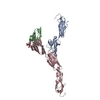
| ||||||||
|---|---|---|---|---|---|---|---|---|---|
| 1 |
| ||||||||
| Unit cell |
| ||||||||
| Components on special symmetry positions |
|

-Components
| #1: Protein | Mass: 33303.145 Da / Num. of mol.: 2 / Fragment: extracellular domains (Ig1-Ig3) / Mutation: N31Q, N262Q, N286Q Source method: isolated from a genetically manipulated source Source: (gene. exp.)  Trichoplusia ni (cabbage looper) / References: UniProt: Q8R464 Trichoplusia ni (cabbage looper) / References: UniProt: Q8R464#2: Protein | | Mass: 15998.754 Da / Num. of mol.: 1 / Fragment: Ig domain Source method: isolated from a genetically manipulated source Source: (gene. exp.)  #3: Polysaccharide | 2-acetamido-2-deoxy-beta-D-glucopyranose-(1-4)-[alpha-L-fucopyranose-(1-6)]2-acetamido-2-deoxy-beta- ...2-acetamido-2-deoxy-beta-D-glucopyranose-(1-4)-[alpha-L-fucopyranose-(1-6)]2-acetamido-2-deoxy-beta-D-glucopyranose | Source method: isolated from a genetically manipulated source Has protein modification | Y | |
|---|
-Experimental details
-Experiment
| Experiment | Method: X-RAY DIFFRACTION / Number of used crystals: 1 |
|---|
- Sample preparation
| Crystal | Density Matthews: 3.98 Å3/Da / Density % sol: 75.78 % / Description: hexagonal prisms-like crystals |
|---|---|
| Crystal grow | Temperature: 291 K / Method: vapor diffusion, sitting drop / pH: 7.5 Details: 0.2M calcium acetate, 0.1M HEPES Sodium Salt ,pH7.5, 10% PEG 8000 |
-Data collection
| Diffraction | Mean temperature: 100 K |
|---|---|
| Diffraction source | Source: SYNCHROTRON / Site: SSRF / Beamline: BL17U1 / Wavelength: 0.97776 Å |
| Detector | Type: ADSC QUANTUM 315r / Detector: CCD / Date: Oct 9, 2016 |
| Radiation | Protocol: SINGLE WAVELENGTH / Monochromatic (M) / Laue (L): M / Scattering type: x-ray |
| Radiation wavelength | Wavelength: 0.97776 Å / Relative weight: 1 |
| Reflection | Resolution: 3.29→49.836 Å / Num. obs: 19407 / % possible obs: 97.19 % / Observed criterion σ(I): -3 / Redundancy: 5.68 % / Biso Wilson estimate: 64.79 Å2 / CC1/2: 0.99 / Rmerge(I) obs: 0.224 / Χ2: 0.98 / Net I/σ(I): 6.96 |
| Reflection shell | Resolution: 3.29→3.49 Å / Redundancy: 5.74 % / Rmerge(I) obs: 1.074 / Mean I/σ(I) obs: 1.64 / Num. unique obs: 3096 / CC1/2: 0.613 / Χ2: 0.89 / % possible all: 97.6 |
- Processing
| Software |
| ||||||||||||||||||||||||||||||||||||||||||||||||||||||||
|---|---|---|---|---|---|---|---|---|---|---|---|---|---|---|---|---|---|---|---|---|---|---|---|---|---|---|---|---|---|---|---|---|---|---|---|---|---|---|---|---|---|---|---|---|---|---|---|---|---|---|---|---|---|---|---|---|---|
| Refinement | Method to determine structure: MOLECULAR REPLACEMENT Starting model: 1Z9M Resolution: 3.29→49.84 Å / SU ML: 0.56 / Cross valid method: THROUGHOUT / σ(F): 1.34 / Phase error: 33.49
| ||||||||||||||||||||||||||||||||||||||||||||||||||||||||
| Displacement parameters | Biso mean: 65.7 Å2 | ||||||||||||||||||||||||||||||||||||||||||||||||||||||||
| Refinement step | Cycle: LAST / Resolution: 3.29→49.84 Å
| ||||||||||||||||||||||||||||||||||||||||||||||||||||||||
| Refine LS restraints |
| ||||||||||||||||||||||||||||||||||||||||||||||||||||||||
| LS refinement shell |
|
