 Movie
Movie Controller

[English] 日本語
 Yorodumi
- EMDB-7032: Global architecture of the HIV-1 reverse transcriptase initiation... -
+ Open data

- Basic information
| Entry | Database: EMDB / ID: EMD-7032 | |||||||||
|---|---|---|---|---|---|---|---|---|---|---|
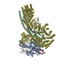
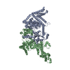
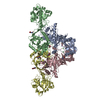
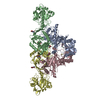
| Title | Global architecture of the HIV-1 reverse transcriptase initiation complex | |||||||||
 Map data Map data | RTIC global architecture | |||||||||
 Sample Sample |
| |||||||||
| Biological species |   Human immunodeficiency virus 1 / Human immunodeficiency virus 1 /  Human (human) / Homo sapiens (human) Human (human) / Homo sapiens (human) | |||||||||
| Method | single particle reconstruction / cryo EM / Resolution: 8.0 Å | |||||||||
 Authors Authors | Larsen KP / Mathiharan YK / Chen DH / Puglisi JD / Skiniotis G / Puglisi EV | |||||||||
| Funding support |  United States, 1 items United States, 1 items
| |||||||||
 Citation Citation | Journal: Nature / Year: 2018 Title: Architecture of an HIV-1 reverse transcriptase initiation complex. Authors: Kevin P Larsen / Yamuna Kalyani Mathiharan / Kalli Kappel / Aaron T Coey / Dong-Hua Chen / Daniel Barrero / Lauren Madigan / Joseph D Puglisi / Georgios Skiniotis / Elisabetta Viani Puglisi / Abstract: Reverse transcription of the HIV-1 RNA genome into double-stranded DNA is a central step in viral infection and a common target of antiretroviral drugs . The reaction is catalysed by viral reverse ...Reverse transcription of the HIV-1 RNA genome into double-stranded DNA is a central step in viral infection and a common target of antiretroviral drugs . The reaction is catalysed by viral reverse transcriptase (RT) that is packaged in an infectious virion with two copies of viral genomic RNA each bound to host lysine 3 transfer RNA (tRNA), which acts as a primer for initiation of reverse transcription. Upon viral entry into cells, initiation is slow and non-processive compared to elongation. Despite extensive efforts, the structural basis of RT function during initiation has remained a mystery. Here we use cryo-electron microscopy to determine a three-dimensional structure of an HIV-1 RT initiation complex. In our structure, RT is in an inactive polymerase conformation with open fingers and thumb and with the nucleic acid primer-template complex shifted away from the active site. The primer binding site (PBS) helix formed between tRNA and HIV-1 RNA lies in the cleft of RT and is extended by additional pairing interactions. The 5' end of the tRNA refolds and stacks on the PBS to create a long helical structure, while the remaining viral RNA forms two helical stems positioned above the RT active site, with a linker that connects these helices to the RNase H region of the PBS. Our results illustrate how RNA structure in the initiation complex alters RT conformation to decrease activity, highlighting a potential target for drug action. | |||||||||
| History |
|
- Structure visualization
| Movie |
 Movie viewer Movie viewer |
|---|---|
| Structure viewer | EM map: SurfViewMolmilJmol/JSmol |
| Supplemental images |

- Downloads & links
-EMDB archive
| Map data | emd_7032.map.gz | 78.1 MB | EMDB map data format | |
|---|---|---|---|---|
| Header (meta data) | emd-7032-v30.xmlemd-7032.xml | 19.3 KB 19.3 KB | Display Display | EMDB header |
| Images | 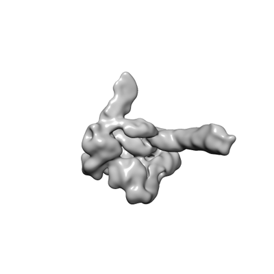 emd_7032.png emd_7032.png | 19.4 KB | ||
| Archive directory |  http://ftp.pdbj.org/pub/emdb/structures/EMD-7032ftp://ftp.pdbj.org/pub/emdb/structures/EMD-7032 http://ftp.pdbj.org/pub/emdb/structures/EMD-7032ftp://ftp.pdbj.org/pub/emdb/structures/EMD-7032 | HTTPS FTP |
-Related structure data


-Links
| EMDB pages | EMDB (EBI/PDBe) / EMDataResource |
|---|
-Map
| File | Download / File: emd_7032.map.gz / Format: CCP4 / Size: 83.7 MB / Type: IMAGE STORED AS FLOATING POINT NUMBER (4 BYTES) | ||||||||||||||||||||||||||||||||||||||||||||||||||||||||||||||||||||
|---|---|---|---|---|---|---|---|---|---|---|---|---|---|---|---|---|---|---|---|---|---|---|---|---|---|---|---|---|---|---|---|---|---|---|---|---|---|---|---|---|---|---|---|---|---|---|---|---|---|---|---|---|---|---|---|---|---|---|---|---|---|---|---|---|---|---|---|---|---|
| Annotation | RTIC global architecture | ||||||||||||||||||||||||||||||||||||||||||||||||||||||||||||||||||||
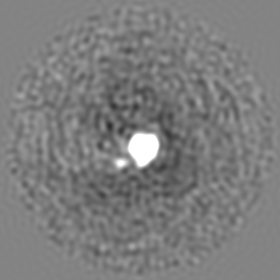
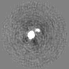
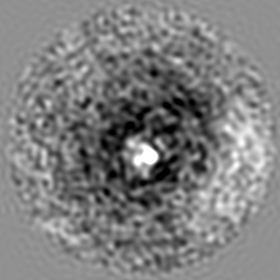
| Projections & slices | Image control
Images are generated by Spider. | ||||||||||||||||||||||||||||||||||||||||||||||||||||||||||||||||||||
| Voxel size | X=Y=Z: 1 Å | ||||||||||||||||||||||||||||||||||||||||||||||||||||||||||||||||||||
| Density |
| ||||||||||||||||||||||||||||||||||||||||||||||||||||||||||||||||||||
| Symmetry | Space group: 1 | ||||||||||||||||||||||||||||||||||||||||||||||||||||||||||||||||||||
| Details | EMDB XML:
CCP4 map header:
| ||||||||||||||||||||||||||||||||||||||||||||||||||||||||||||||||||||
 Z (Sec.)
 Y (Row.)
 X (Col.)


-Supplemental data
- Sample components
-Entire : HIV-1 reverse transcriptase initiation complex
| Entire | Name: HIV-1 reverse transcriptase initiation complex |
|---|---|
| Components |
|
-Supramolecule #1: HIV-1 reverse transcriptase initiation complex
| Supramolecule | Name: HIV-1 reverse transcriptase initiation complex / type: complex / ID: 1 / Parent: 0 / Macromolecule list: all |
|---|---|
| Molecular weight | Theoretical: 175 KDa |
-Supramolecule #2: HIV-1 reverse transcriptase
| Supramolecule | Name: HIV-1 reverse transcriptase / type: complex / ID: 2 / Parent: 1 / Macromolecule list: #1-#2 Details: A cysteine mutation for crosslinking was introduced into helix H of p66 (Q258C). The protein used in this study also had the C280S mutation, introduced in prior structural work and the E478Q ...Details: A cysteine mutation for crosslinking was introduced into helix H of p66 (Q258C). The protein used in this study also had the C280S mutation, introduced in prior structural work and the E478Q mutation, introduced to eliminate RNase H activity. |
|---|---|
| Source (natural) | Organism: Human immunodeficiency virus 1 |
| Recombinant expression | Organism:  |
| Molecular weight | Theoretical: 117 KDa |
-Supramolecule #3: tRNA lysine3 primer
| Supramolecule | Name: tRNA lysine3 primer / type: complex / ID: 3 / Parent: 1 / Macromolecule list: #3 Details: Chemically synthesized and extended tRNA lysine3 primer. Modified nucleotide containing a N2-cystamine was placed at position 71. The tRNA primer has been extended by one ddCTP, bringing its ...Details: Chemically synthesized and extended tRNA lysine3 primer. Modified nucleotide containing a N2-cystamine was placed at position 71. The tRNA primer has been extended by one ddCTP, bringing its total length in the full complex to 77 nucleotides. |
|---|---|
| Source (natural) | Organism: Human (human) |
| Molecular weight | Theoretical: 25 KDa |
-Supramolecule #4: HIV-1 RNA genome fragment
| Supramolecule | Name: HIV-1 RNA genome fragment / type: complex / ID: 4 / Parent: 1 / Macromolecule list: #4 Details: HIV-1 RNA genome fragment of 101 nucleotides in length. Contains the primer binding site (PBS), primer activation signal (PAS), A-rich loop, and C-rich region. |
|---|---|
| Source (natural) | Organism: Human immunodeficiency virus 1 |
| Molecular weight | Theoretical: 33 KDa |
-Macromolecule #1: HIV-1 reverse transcriptase: p66 subunit
| Macromolecule | Name: HIV-1 reverse transcriptase: p66 subunit / type: protein_or_peptide / ID: 1 / Enantiomer: LEVO |
|---|---|
| Source (natural) | Organism: Human immunodeficiency virus 1 |
| Recombinant expression | Organism: |
| Sequence | String: MVPISPIETV PVKLKPGMDG PKVKQWPLTE EKIKALVEIC TEMEKEGKIS KIGPENPYNT PVFAIKKKDS TKWRKLVDFR ELNKRTQDFW EVQLGIPHPA GLKKKKSVTV LDVGDAYFSV PLDEDFRKYT AFTIPSINNE TPGIRYQYNV LPQGWKGSPA IFQSSMTKIL ...String: MVPISPIETV PVKLKPGMDG PKVKQWPLTE EKIKALVEIC TEMEKEGKIS KIGPENPYNT PVFAIKKKDS TKWRKLVDFR ELNKRTQDFW EVQLGIPHPA GLKKKKSVTV LDVGDAYFSV PLDEDFRKYT AFTIPSINNE TPGIRYQYNV LPQGWKGSPA IFQSSMTKIL EPFKKQNPDI VIYQYMDDLY VGSDLEIGQH RTKIEELRQH LLRWGLTTPD KKHQKEPPFL WMGYELHPDK WTVQPIVLPE KDSWTVNDIC KLVGKLNWAS QIYPGIKVRQ LSKLLRGTKA LTEVIPLTEE AELELAENRE ILKEPVHGVY YDPSKDLIAE IQKQGQGQWT YQIYQEPFKN LKTGKYARMR GAHTNDVKQL TEAVQKITTE SIVIWGKTPK FKLPIQKETW ETWWTEYWQA TWIPEWEFVN TPPLVKLWYQ LEKEPIVGAE TFYVDGAANR ETKLGKAGYV TNKGRQKVVP LTNTTNQKTQ LQAIYLALQD SGLEVNIVTD SQYALGIIQA QPDKSESELV NQIIEQLIKK EKVYLAWVPA HKGIGGNEQV DKLVSAGIRK IL |
-Macromolecule #2: HIV-1 reverse transcriptase: p51 subunit
| Macromolecule | Name: HIV-1 reverse transcriptase: p51 subunit / type: protein_or_peptide / ID: 2 / Enantiomer: LEVO |
|---|---|
| Source (natural) | Organism: Human immunodeficiency virus 1 |
| Recombinant expression | Organism: |
| Sequence | String: MVPISPIETV PVKLKPGMDG PKVKQWPLTE EKIKALVEIC TEMEKEGKIS KIGPENPYNT PVFAIKKKDS TKWRKLVDFR ELNKRTQDFW EVQLGIPHPA GLKKKKSVTV LDVGDAYFSV PLDEDFRKYT AFTIPSINNE TPGIRYQYNV LPQGWKGSPA IFQSSMTKIL ...String: MVPISPIETV PVKLKPGMDG PKVKQWPLTE EKIKALVEIC TEMEKEGKIS KIGPENPYNT PVFAIKKKDS TKWRKLVDFR ELNKRTQDFW EVQLGIPHPA GLKKKKSVTV LDVGDAYFSV PLDEDFRKYT AFTIPSINNE TPGIRYQYNV LPQGWKGSPA IFQSSMTKIL EPFKKQNPDI VIYQYMDDLY VGSDLEIGQH RTKIEELRQH LLRWGLTTPD KKHQKEPPFL WMGYELHPDK WTVQPIVLPE KDSWTVNDIQ KLVGKLNWAS QIYPGIKVRQ LSKLLRGTKA LTEVIPLTEE AELELAENRE ILKEPVHGVY YDPSKDLIAE IQKQGQGQWT YQIYQEPFKN LKTGKYARMR GAHTNDVKQL TEAVQKITTE SIVIWGKTPK FKLPIQKETW ETWWTEYWQA TWIPEWEFVN TPPLVKLWYQ LEKEPIVGAE TF |
-Macromolecule #3: tRNA Lysine3
| Macromolecule | Name: tRNA Lysine3 / type: rna / ID: 3 Details: 77C is ddC (from RT extension); 71G is dG with an N2-cystamine for cross linking to RT-p66 |
|---|---|
| Source (natural) | Organism: Homo sapiens (human) |
| Sequence | String: GCCCGGAUAG CUCAGUCGGU AGAGCAUCAG ACUUUUAAUC UGAGGGUCCA GGGUUCAAGU CCCUGUUCGG GCGCCAC |
-Macromolecule #4: HIV-1 viral RNA genome fragment
| Macromolecule | Name: HIV-1 viral RNA genome fragment / type: rna / ID: 4 |
|---|---|
| Source (natural) | Organism: Human immunodeficiency virus 1 / Strain: NL4.3 |
| Sequence | String: GACUCUGGUA ACUAGAGAUC CCUCAGACCC UUUUAGUCAG UGUGGAAAAU CUCUAGCAGU GGCGCCCGAA CAGGGACUUG AAAGCGAAAG UAAAGCCAGA G |
-Experimental details
-Structure determination
| Method | cryo EM |
|---|---|
 Processing Processing | single particle reconstruction |
| Aggregation state | particle |
-Sample preparation
| Concentration | 5.0 mg/mL | ||||||||||||
|---|---|---|---|---|---|---|---|---|---|---|---|---|---|
| Buffer | pH: 8 Component:
Details: Beta-OG was added just prior to freezing. | ||||||||||||
| Grid | Model: Quantifoil R2/2 / Material: COPPER / Mesh: 200 / Pretreatment - Type: GLOW DISCHARGE | ||||||||||||
| Vitrification | Cryogen name: ETHANE / Chamber humidity: 85 % / Chamber temperature: 292 K / Instrument: FEI VITROBOT MARK IV Details: Blotted for 3.5 sec before plunging into liquid ethane.. | ||||||||||||
| Details | Sample was monodisperse. |
- Electron microscopy
| Microscope | FEI TITAN KRIOS |
|---|---|
| Image recording | Film or detector model: GATAN K2 SUMMIT (4k x 4k) / Detector mode: COUNTING / Digitization - Frames/image: 1-40 / Number real images: 4209 / Average exposure time: 8.0 sec. / Average electron dose: 70.0 e/Å2 |
| Electron beam | Acceleration voltage: 300 kV / Electron source:  FIELD EMISSION GUN FIELD EMISSION GUN |
| Electron optics | Calibrated magnification: 50000 / Illumination mode: FLOOD BEAM / Imaging mode: BRIGHT FIELD / Cs: 2.7 mm / Nominal defocus max: 2.5 µm / Nominal defocus min: 1.3 µm / Nominal magnification: 29000 |
| Sample stage | Specimen holder model: FEI TITAN KRIOS AUTOGRID HOLDER / Cooling holder cryogen: NITROGEN |
| Experimental equipment |  Model: Titan Krios / Image courtesy: FEI Company |
