 Movie
Movie Controller


+ Open data

- Basic information
| Entry | Database: PDB / ID: 2y9z | ||||||
|---|---|---|---|---|---|---|---|
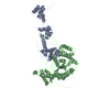
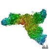
| Title | Chromatin Remodeling Factor ISW1a(del_ATPase) in DNA complex | ||||||
 Components Components |
| ||||||
 Keywords Keywords | TRANSCRIPTION / NUCLEAR PROTEIN-DNA COMPLEX / CHROMATIN REMODELING / NUCLEOSOME REMODELING | ||||||
| Function / homology |  Function and homology information Function and homology informationIsw1b complex / Isw1a complex / Isw1 complex / regulation of transcriptional start site selection at RNA polymerase II promoter / nucleolar chromatin / negative regulation of RNA export from nucleus / negative regulation of IRE1-mediated unfolded protein response / regulation of chromatin organization / rDNA binding / nucleosome array spacer activity ...Isw1b complex / Isw1a complex / Isw1 complex / regulation of transcriptional start site selection at RNA polymerase II promoter / nucleolar chromatin / negative regulation of RNA export from nucleus / negative regulation of IRE1-mediated unfolded protein response / regulation of chromatin organization / rDNA binding / nucleosome array spacer activity / DNA-templated transcription elongation / sister chromatid cohesion / termination of RNA polymerase II transcription / termination of RNA polymerase I transcription / nucleosome binding / helicase activity / mRNA 3'-UTR binding / Hydrolases; Acting on acid anhydrides; Acting on acid anhydrides to facilitate cellular and subcellular movement / heterochromatin formation / response to heat / transcription cis-regulatory region binding / chromatin remodeling / hydrolase activity / chromatin binding / regulation of DNA-templated transcription / chromatin / positive regulation of transcription by RNA polymerase II / ATP binding / nucleus Similarity search - Function | ||||||
| Biological species |  | ||||||
| Method |  X-RAY DIFFRACTION / SYNCHROTRON / MIRAS / Resolution: 3.601 Å X-RAY DIFFRACTION / SYNCHROTRON / MIRAS / Resolution: 3.601 Å | ||||||
 Authors Authors | Yamada, K. / Frouws, T.D. / Angst, B. / Fitzgerald, D.J. / DeLuca, C. / Schimmele, K. / Sargent, D.F. / Richmond, T.J. | ||||||
 Citation Citation | Journal: Nature / Year: 2011 Title: Structure and mechanism of the chromatin remodelling factor ISW1a. Authors: Kazuhiro Yamada / Timothy D Frouws / Brigitte Angst / Daniel J Fitzgerald / Carl DeLuca / Kyoko Schimmele / David F Sargent / Timothy J Richmond /  Abstract: Site-specific recognition of DNA in eukaryotic organisms depends on the arrangement of nucleosomes in chromatin. In the yeast Saccharomyces cerevisiae, ISW1a and related chromatin remodelling factors ...Site-specific recognition of DNA in eukaryotic organisms depends on the arrangement of nucleosomes in chromatin. In the yeast Saccharomyces cerevisiae, ISW1a and related chromatin remodelling factors are implicated in establishing the nucleosome repeat during replication and altering nucleosome position to affect gene activity. Here we have solved the crystal structures of S. cerevisiae ISW1a lacking its ATPase domain both alone and with DNA bound at resolutions of 3.25 Å and 3.60 Å, respectively, and we have visualized two different nucleosome-containing remodelling complexes using cryo-electron microscopy. The composite X-ray and electron microscopy structures combined with site-directed photocrosslinking analyses of these complexes suggest that ISW1a uses a dinucleosome substrate for chromatin remodelling. Results from a remodelling assay corroborate the dinucleosome model. We show how a chromatin remodelling factor could set the spacing between two adjacent nucleosomes acting as a 'protein ruler'. | ||||||
| History |
| ||||||
| Remark 650 | HELIX DETERMINATION METHOD: AUTHOR PROVIDED. | ||||||
| Remark 700 | SHEET DETERMINATION METHOD: AUTHOR PROVIDED. |
- Structure visualization
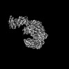
| Structure viewer | Molecule: MolmilJmol/JSmol |
|---|
- Downloads & links
-Download
| PDBx/mmCIF format | 2y9z.cif.gz | 507.6 KB | Display | PDBx/mmCIF format |
|---|---|---|---|---|
| PDB format | pdb2y9z.ent.gz | 413.3 KB | Display | PDB format |
| PDBx/mmJSON format | 2y9z.json.gz | Tree view | PDBx/mmJSON format | |
| Others |  Other downloads Other downloads |
-Validation report
| Arichive directory | https://data.pdbj.org/pub/pdb/validation_reports/y9/2y9zftp://data.pdbj.org/pub/pdb/validation_reports/y9/2y9z | HTTPS FTP |
|---|
-Related structure data


-Links
 PDBj
PDBj


- Assembly
| Deposited unit | 
| ||||||||
|---|---|---|---|---|---|---|---|---|---|
| 1 |
| ||||||||
| Unit cell |
|
-Components
| #1: Protein | Mass: 44327.895 Da / Num. of mol.: 1 / Fragment: HAND, SANT, SLIDE DOMAINS, RESIDUES 763-1129 Source method: isolated from a genetically manipulated source Source: (gene. exp.) Plasmid: MULTIBAC / Production host:   SPODOPTERA FRUGIPERDA (fall armyworm) / Strain (production host): SF21 / References: UniProt: P38144 SPODOPTERA FRUGIPERDA (fall armyworm) / Strain (production host): SF21 / References: UniProt: P38144 |
|---|---|
| #2: Protein | Mass: 72422.539 Da / Num. of mol.: 1 Fragment: CORE DOMAIN CONTAINING CLB AND HLB SUBDOMAINS, RESIDUES 127-749 Source method: isolated from a genetically manipulated source Source: (gene. exp.) Plasmid: MULTIBAC / Production host: SPODOPTERA FRUGIPERDA (fall armyworm) / Strain (production host): SF21 / References: UniProt: P43596 |
| #3: DNA chain | Mass: 14784.487 Da / Num. of mol.: 4 / Source method: obtained synthetically / Details: 48 BASES, C-D I-DNA DUPLEX, E-F E-DNA DUPLEX / Source: (synth.) |
-Experimental details
-Experiment
| Experiment | Method: X-RAY DIFFRACTION / Number of used crystals: 8 |
|---|
- Sample preparation
| Crystal | Density Matthews: 6.7 Å3/Da / Density % sol: 83 % / Description: NONE |
|---|---|
| Crystal grow | pH: 7 Details: 50MM BISTRIS(PH7.0), 150MM NACITRATE, 30%(W/V) PEG5000 |
-Data collection
| Diffraction | Mean temperature: 100 K |
|---|---|
| Diffraction source | Source: SYNCHROTRON / Site: SLS / Beamline: X06SA / Wavelength: 0.9999 |
| Detector | Type: PSI PILATUS 6M / Detector: PIXEL / Date: May 18, 2009 |
| Radiation | Monochromator: LN2 COOLED FIXED-EXIT SI(111) MONOCHROMATOR / Protocol: SINGLE WAVELENGTH / Monochromatic (M) / Laue (L): M / Scattering type: x-ray |
| Radiation wavelength | Wavelength: 0.9999 Å / Relative weight: 1 |
| Reflection | Resolution: 3.6→30 Å / Num. obs: 53279 / % possible obs: 99.8 % / Observed criterion σ(I): 2 / Redundancy: 7.4 % / Biso Wilson estimate: 118.01 Å2 / Rmerge(I) obs: 0.07 / Net I/σ(I): 5.9 |
| Reflection shell | Resolution: 3.6→3.67 Å / Redundancy: 7.3 % / Rmerge(I) obs: 0.36 / Mean I/σ(I) obs: 2 / % possible all: 99.8 |
- Processing
| Software |
| ||||||||||||||||||||||||||||||||||||||||||||||||||||||||||||||||||||||||||||||||||||||||||||||||||||||||||||||||||||||||||||||||||||||||||||||||||||||
|---|---|---|---|---|---|---|---|---|---|---|---|---|---|---|---|---|---|---|---|---|---|---|---|---|---|---|---|---|---|---|---|---|---|---|---|---|---|---|---|---|---|---|---|---|---|---|---|---|---|---|---|---|---|---|---|---|---|---|---|---|---|---|---|---|---|---|---|---|---|---|---|---|---|---|---|---|---|---|---|---|---|---|---|---|---|---|---|---|---|---|---|---|---|---|---|---|---|---|---|---|---|---|---|---|---|---|---|---|---|---|---|---|---|---|---|---|---|---|---|---|---|---|---|---|---|---|---|---|---|---|---|---|---|---|---|---|---|---|---|---|---|---|---|---|---|---|---|---|---|---|---|
| Refinement | Method to determine structure: MIRAS Starting model: NONE Resolution: 3.601→29.955 Å / SU ML: 0.47 / σ(F): 1.32 / Phase error: 32.1 / Stereochemistry target values: MLHL Details: CHAIN A RESIDUES N-TERMINUS TO 788 ARE DISORDERED. CHAIN A RESIDUES 1076 TO C-TERMINUS ARE DISORDERED. CHAIN B RESIDUES N-TERMINUS TO 133 ARE DISORDERED. CHAIN B RESIDUES 658 TO 677 ARE ...Details: CHAIN A RESIDUES N-TERMINUS TO 788 ARE DISORDERED. CHAIN A RESIDUES 1076 TO C-TERMINUS ARE DISORDERED. CHAIN B RESIDUES N-TERMINUS TO 133 ARE DISORDERED. CHAIN B RESIDUES 658 TO 677 ARE DISORDERED. CHAIN B RESIDUES 749 TO C-TERMINUS ARE DISORDERED. CHAIN C BASES 1 TO 7 ARE DISORDERED. CHAIN C BASES 39 TO 48 ARE DISORDERED. CHAIN D BASES 1 TO 10 ARE DISORDERED. CHAIN D BASES 42 TO 48 ARE DISORDERED. CHAIN E BASES 1 TO 24 ARE DISORDERED. CHAIN E BASE 48 IS DISORDERED. CHAIN F BASE 1 IS DISORDERED. CHAIN F BASES 25 TO 48 ARE DISORDERED.
| ||||||||||||||||||||||||||||||||||||||||||||||||||||||||||||||||||||||||||||||||||||||||||||||||||||||||||||||||||||||||||||||||||||||||||||||||||||||
| Solvent computation | Shrinkage radii: 0.9 Å / VDW probe radii: 1.11 Å / Solvent model: FLAT BULK SOLVENT MODEL / Bsol: 95.002 Å2 / ksol: 0.313 e/Å3 | ||||||||||||||||||||||||||||||||||||||||||||||||||||||||||||||||||||||||||||||||||||||||||||||||||||||||||||||||||||||||||||||||||||||||||||||||||||||
| Displacement parameters | Biso mean: 172.3 Å2
| ||||||||||||||||||||||||||||||||||||||||||||||||||||||||||||||||||||||||||||||||||||||||||||||||||||||||||||||||||||||||||||||||||||||||||||||||||||||
| Refinement step | Cycle: LAST / Resolution: 3.601→29.955 Å
| ||||||||||||||||||||||||||||||||||||||||||||||||||||||||||||||||||||||||||||||||||||||||||||||||||||||||||||||||||||||||||||||||||||||||||||||||||||||
| Refine LS restraints |
| ||||||||||||||||||||||||||||||||||||||||||||||||||||||||||||||||||||||||||||||||||||||||||||||||||||||||||||||||||||||||||||||||||||||||||||||||||||||
| LS refinement shell |
| ||||||||||||||||||||||||||||||||||||||||||||||||||||||||||||||||||||||||||||||||||||||||||||||||||||||||||||||||||||||||||||||||||||||||||||||||||||||
| Refinement TLS params. | Method: refined / Refine-ID: X-RAY DIFFRACTION
| ||||||||||||||||||||||||||||||||||||||||||||||||||||||||||||||||||||||||||||||||||||||||||||||||||||||||||||||||||||||||||||||||||||||||||||||||||||||
| Refinement TLS group |
|
