Movie
Movie Controller

[English] 日本語
 Yorodumi
- EMDB-1877: CryoEM structure of the chromatin remodelling factor ISW1a bound ... -
+ Open data

- Basic information
| Entry | Database: EMDB / ID: EMD-1877 | |||||||||
|---|---|---|---|---|---|---|---|---|---|---|
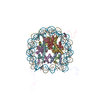
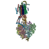
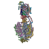
| Title | CryoEM structure of the chromatin remodelling factor ISW1a bound to a mononucleosome (45N29) | |||||||||
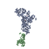
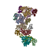
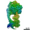
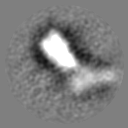
 Map data Map data | Surface rendering of 45N29-ISW1a (delta ATPase) | |||||||||
 Sample Sample |
| |||||||||
 Keywords Keywords | Nucleosome / chromatin remodelling factor / ISW1a / ISWI | |||||||||
| Biological species |  | |||||||||
| Method | single particle reconstruction / cryo EM / Resolution: 22.0 Å | |||||||||
 Authors Authors | Frouws TD / Richmond TJ | |||||||||
 Citation Citation | Journal: Nature / Year: 2011 Title: Structure and mechanism of the chromatin remodelling factor ISW1a. Authors: Kazuhiro Yamada / Timothy D Frouws / Brigitte Angst / Daniel J Fitzgerald / Carl DeLuca / Kyoko Schimmele / David F Sargent / Timothy J Richmond /  Abstract: Site-specific recognition of DNA in eukaryotic organisms depends on the arrangement of nucleosomes in chromatin. In the yeast Saccharomyces cerevisiae, ISW1a and related chromatin remodelling factors ...Site-specific recognition of DNA in eukaryotic organisms depends on the arrangement of nucleosomes in chromatin. In the yeast Saccharomyces cerevisiae, ISW1a and related chromatin remodelling factors are implicated in establishing the nucleosome repeat during replication and altering nucleosome position to affect gene activity. Here we have solved the crystal structures of S. cerevisiae ISW1a lacking its ATPase domain both alone and with DNA bound at resolutions of 3.25 Å and 3.60 Å, respectively, and we have visualized two different nucleosome-containing remodelling complexes using cryo-electron microscopy. The composite X-ray and electron microscopy structures combined with site-directed photocrosslinking analyses of these complexes suggest that ISW1a uses a dinucleosome substrate for chromatin remodelling. Results from a remodelling assay corroborate the dinucleosome model. We show how a chromatin remodelling factor could set the spacing between two adjacent nucleosomes acting as a 'protein ruler'. | |||||||||
| History |
|
- Structure visualization
| Movie |
 Movie viewer Movie viewer |
|---|---|
| Structure viewer | EM map: SurfViewMolmilJmol/JSmol |
| Supplemental images |

- Downloads & links
-EMDB archive
| Map data | emd_1877.map.gz | 3.3 MB | EMDB map data format | |
|---|---|---|---|---|
| Header (meta data) | emd-1877-v30.xmlemd-1877.xml | 10.8 KB 10.8 KB | Display Display | EMDB header |
| Images |  EMD-1877image.png EMD-1877image.png | 100.1 KB | ||
| Archive directory |  http://ftp.pdbj.org/pub/emdb/structures/EMD-1877ftp://ftp.pdbj.org/pub/emdb/structures/EMD-1877 http://ftp.pdbj.org/pub/emdb/structures/EMD-1877ftp://ftp.pdbj.org/pub/emdb/structures/EMD-1877 | HTTPS FTP |
-Related structure data

-Links
| EMDB pages | EMDB (EBI/PDBe) / EMDataResource |
|---|
-Map
| File | Download / File: emd_1877.map.gz / Format: CCP4 / Size: 7.8 MB / Type: IMAGE STORED AS FLOATING POINT NUMBER (4 BYTES) | ||||||||||||||||||||||||||||||||||||||||||||||||||||||||||||||||||||
|---|---|---|---|---|---|---|---|---|---|---|---|---|---|---|---|---|---|---|---|---|---|---|---|---|---|---|---|---|---|---|---|---|---|---|---|---|---|---|---|---|---|---|---|---|---|---|---|---|---|---|---|---|---|---|---|---|---|---|---|---|---|---|---|---|---|---|---|---|---|
| Annotation | Surface rendering of 45N29-ISW1a (delta ATPase) | ||||||||||||||||||||||||||||||||||||||||||||||||||||||||||||||||||||
| Projections & slices | Image control
Images are generated by Spider. | ||||||||||||||||||||||||||||||||||||||||||||||||||||||||||||||||||||
| Voxel size | X=Y=Z: 2.7 Å | ||||||||||||||||||||||||||||||||||||||||||||||||||||||||||||||||||||
| Density |
| ||||||||||||||||||||||||||||||||||||||||||||||||||||||||||||||||||||
| Symmetry | Space group: 1 | ||||||||||||||||||||||||||||||||||||||||||||||||||||||||||||||||||||
| Details | EMDB XML:
CCP4 map header:
| ||||||||||||||||||||||||||||||||||||||||||||||||||||||||||||||||||||
 Z (Sec.)
 Y (Row.)
 X (Col.)


-Supplemental data
- Sample components
-Entire : Chromatin remodelling factor ISW1a (delta ATPase) bound to a mono...
| Entire | Name: Chromatin remodelling factor ISW1a (delta ATPase) bound to a mononucleosome (with 45 and 29 bp extensions). |
|---|---|
| Components |
|
-Supramolecule #1000: Chromatin remodelling factor ISW1a (delta ATPase) bound to a mono...
| Supramolecule | Name: Chromatin remodelling factor ISW1a (delta ATPase) bound to a mononucleosome (with 45 and 29 bp extensions). type: sample / ID: 1000 / Oligomeric state: Monomer / Number unique components: 2 |
|---|---|
| Molecular weight | Theoretical: 380 KDa |
-Macromolecule #1: Nucleosome
| Macromolecule | Name: Nucleosome / type: protein_or_peptide / ID: 1 / Name.synonym: Nucleosome Details: Nucleosomes consist of recombinant xenopus histones H2A,H2B,H3,H4, reconstituted onto '601' DNA with 45 and 29 bp extensions. Recombinant expression: Yes |
|---|---|
| Source (natural) | Organism: |
| Molecular weight | Theoretical: 256 MDa |
| Recombinant expression | Organism:  |
-Macromolecule #2: ISW1a
| Macromolecule | Name: ISW1a / type: protein_or_peptide / ID: 2 / Name.synonym: Remodelling factor Details: Recombinant yeast ISW1a (delta ATPase) expressed with MultiBac system Recombinant expression: Yes |
|---|---|
| Source (natural) | Organism: |
| Molecular weight | Theoretical: 120 MDa |
| Recombinant expression | Organism:   Spodoptera frugiperda (fall armyworm) Spodoptera frugiperda (fall armyworm) |
-Experimental details
-Structure determination
| Method | cryo EM |
|---|---|
 Processing Processing | single particle reconstruction |
| Aggregation state | particle |
-Sample preparation
| Concentration | 3.5 mg/mL |
|---|---|
| Buffer | pH: 7.5 / Details: 10mM TrisCl, 10mM KCl, 0.1mM EDTA |
| Grid | Details: C-flat 224 |
| Vitrification | Cryogen name: ETHANE / Chamber humidity: 90 % / Chamber temperature: 100 K / Instrument: FEI VITROBOT MARK II / Details: Vitrification instrument: Vitribot MKII / Method: 0 offset, 4 sec blot, 1 sec drain |
- Electron microscopy
| Microscope | FEI TECNAI F20 |
|---|---|
| Temperature | Average: 100 K |
| Specialist optics | Energy filter - Name: Gatan |
| Details | Low dose |
| Image recording | Category: CCD / Film or detector model: GENERIC GATAN / Digitization - Sampling interval: 14 µm / Average electron dose: 20 e/Å2 / Bits/pixel: 16 |
| Electron beam | Acceleration voltage: 200 kV / Electron source:  FIELD EMISSION GUN FIELD EMISSION GUN |
| Electron optics | Calibrated magnification: 51606 / Illumination mode: FLOOD BEAM / Imaging mode: BRIGHT FIELD / Cs: 2.0 mm / Nominal defocus max: 4.5 µm / Nominal defocus min: 1.5 µm / Nominal magnification: 80000 |
| Sample stage | Specimen holder: Eucentric / Specimen holder model: GATAN LIQUID NITROGEN |
| Experimental equipment |  Model: Tecnai F20 / Image courtesy: FEI Company |
+Image processing
-Atomic model buiding 1
| Initial model | PDB ID: |
|---|---|
| Software | Name: Chimera |
| Details | Manually fitted in Chimera, and missing DNA segments were built in. |
| Refinement | Space: REAL / Protocol: RIGID BODY FIT |