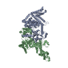
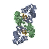
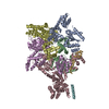
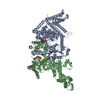
[English] 日本語
Yorodumi
- PDB-4ft2: crystal structure of Zea mays ZMET2 in complex H3(1-15)K9me2 pept... -

+
Open data


ID or keywords:

Loading...

-
Basic information

Entry
Database: PDB / ID: 4ft2
Titlecrystal structure of Zea mays ZMET2 in complex H3(1-15)K9me2 peptide and SAH
Components
  • DNA (cytosine-5)-methyltransferase 1
  • H3 peptide
KeywordsTRANSFERASE / chromodomain / BAH domain / DNA methyltransferase domain / DNA methyltransferase / H3K9me2 binding / methylation
Function / homology
Function and homology information


: / DNA (cytosine-5-)-methyltransferase / DNA (cytosine-5-)-methyltransferase activity / Chromatin modifying enzymes / epigenetic regulation of gene expression / telomere organization / RNA Polymerase I Promoter Opening / Interleukin-7 signaling / Assembly of the ORC complex at the origin of replication / DNA methylation ...: / DNA (cytosine-5-)-methyltransferase / DNA (cytosine-5-)-methyltransferase activity / Chromatin modifying enzymes / epigenetic regulation of gene expression / telomere organization / RNA Polymerase I Promoter Opening / Interleukin-7 signaling / Assembly of the ORC complex at the origin of replication / DNA methylation / Condensation of Prophase Chromosomes / HCMV Late Events / Chromatin modifications during the maternal to zygotic transition (MZT) / ERCC6 (CSB) and EHMT2 (G9a) positively regulate rRNA expression / SIRT1 negatively regulates rRNA expression / PRC2 methylates histones and DNA / Defective pyroptosis / HDACs deacetylate histones / RNA Polymerase I Promoter Escape / Transcriptional regulation by small RNAs / Formation of the beta-catenin:TCF transactivating complex / RUNX1 regulates genes involved in megakaryocyte differentiation and platelet function / Activated PKN1 stimulates transcription of AR (androgen receptor) regulated genes KLK2 and KLK3 / NoRC negatively regulates rRNA expression / B-WICH complex positively regulates rRNA expression / HDMs demethylate histones / PKMTs methylate histone lysines / RMTs methylate histone arginines / Meiotic recombination / Pre-NOTCH Transcription and Translation / nucleosome assembly / Activation of anterior HOX genes in hindbrain development during early embryogenesis / HCMV Early Events / Transcriptional regulation of granulopoiesis / structural constituent of chromatin / nucleosome / gene expression / RUNX1 regulates transcription of genes involved in differentiation of HSCs / chromatin organization / Factors involved in megakaryocyte development and platelet production / HATs acetylate histones / Senescence-Associated Secretory Phenotype (SASP) / Oxidative Stress Induced Senescence / Estrogen-dependent gene expression / cadherin binding / Amyloid fiber formation / protein heterodimerization activity / chromatin binding / protein-containing complex / DNA binding / extracellular exosome / extracellular region / nucleoplasm / membrane / nucleus
Similarity search - Function
Bromo adjacent homology (BAH) domain / DNA Methylase, subunit A, domain 2 / DNA Methylase; Chain A, domain 2 / DNA methylase, C-5 cytosine-specific, active site / C-5 cytosine-specific DNA methylases active site. / C-5 cytosine-specific DNA methylase (Dnmt) domain profile. / C-5 cytosine methyltransferase / C-5 cytosine-specific DNA methylase / Chromo domain / Chromo (CHRromatin Organisation MOdifier) domain ...Bromo adjacent homology (BAH) domain / DNA Methylase, subunit A, domain 2 / DNA Methylase; Chain A, domain 2 / DNA methylase, C-5 cytosine-specific, active site / C-5 cytosine-specific DNA methylases active site. / C-5 cytosine-specific DNA methylase (Dnmt) domain profile. / C-5 cytosine methyltransferase / C-5 cytosine-specific DNA methylase / Chromo domain / Chromo (CHRromatin Organisation MOdifier) domain / Chromo and chromo shadow domain profile. / Bromo adjacent homology (BAH) domain superfamily / Bromo adjacent homology domain / Bromo adjacent homology (BAH) domain / BAH domain / BAH domain profile. / Chromo/chromo shadow domain / Chromatin organization modifier domain / Chromo-like domain superfamily / Vaccinia Virus protein VP39 / Histone H3 signature 1. / Histone H3 signature 2. / Histone H3 / Histone H3/CENP-A / Histone H2A/H2B/H3 / Core histone H2A/H2B/H3/H4 / SH3 type barrels. / Histone-fold / Roll / S-adenosyl-L-methionine-dependent methyltransferase superfamily / Alpha-Beta Complex / Rossmann fold / 3-Layer(aba) Sandwich / Mainly Beta / Alpha Beta
Similarity search - Domain/homology
S-ADENOSYL-L-HOMOCYSTEINE / Histone H3.1 / DNA (cytosine-5)-methyltransferase 1
Similarity search - Component
Biological speciesZea mays (maize)
MethodX-RAY DIFFRACTION / SYNCHROTRON / MOLECULAR REPLACEMENT / Resolution: 3.2 Å
AuthorsDu, J. / Patel, D.J.
CitationJournal: Cell(Cambridge,Mass.) / Year: 2012
Title: Dual Binding of Chromomethylase Domains to H3K9me2-Containing Nucleosomes Directs DNA Methylation in Plants.
Authors: Du, J. / Zhong, X. / Bernatavichute, Y.V. / Stroud, H. / Feng, S. / Caro, E. / Vashisht, A.A. / Terragni, J. / Chin, H.G. / Tu, A. / Hetzel, J. / Wohlschlegel, J.A. / Pradhan, S. / Patel, D.J. / Jacobsen, S.E.
History
DepositionJun 27, 2012Deposition site: RCSB / Processing site: RCSB
Revision 1.0Oct 17, 2012Provider: repository / Type: Initial release
Revision 1.1Sep 13, 2023Group: Data collection / Database references ...Data collection / Database references / Derived calculations / Refinement description
Category: chem_comp_atom / chem_comp_bond ...chem_comp_atom / chem_comp_bond / database_2 / pdbx_initial_refinement_model / struct_conn / struct_ref_seq_dif / struct_site
Item: _database_2.pdbx_DOI / _database_2.pdbx_database_accession ..._database_2.pdbx_DOI / _database_2.pdbx_database_accession / _struct_conn.pdbx_leaving_atom_flag / _struct_ref_seq_dif.details / _struct_site.pdbx_auth_asym_id / _struct_site.pdbx_auth_comp_id / _struct_site.pdbx_auth_seq_id

-
Structure visualization

Structure viewerMolecule:
MolmilJmol/JSmol

Downloads & links

-
Assembly

Deposited unit
A: DNA (cytosine-5)-methyltransferase 1
B: DNA (cytosine-5)-methyltransferase 1
P: H3 peptide
hetero molecules


Theoretical massNumber of molelcules
Total (without water)177,5455
Polymers176,7763
Non-polymers7692
Water0
1


  • Idetical with deposited unit
  • defined by author
TypeNameSymmetry operationNumber
identity operation1_555x,y,z1
Unit cell
Length a, b, c (Å)64.861, 88.858, 113.501
Angle α, β, γ (deg.)93.15, 95.72, 110.70
Int Tables number1
Space group name H-MP1

-
Components

#1: Protein DNA (cytosine-5)-methyltransferase 1 / Chromomethylase 1 / DNA cytosine methyltransferase MET2a / Zea methyltransferase2 / Zmet2


Mass: 87591.828 Da / Num. of mol.: 2 / Fragment: UNP residues 130-912
Source method: isolated from a genetically manipulated source
Source: (gene. exp.) Zea mays (maize) / Gene: MET2A, ZMET2 / Plasmid: pET-Sumo / Production host: Escherichia coli (E. coli) / Strain (production host): BL21(DE3) RIL
References: UniProt: Q9AXT8, DNA (cytosine-5-)-methyltransferase
#2: Protein/peptide H3 peptide


Mass: 1592.843 Da / Num. of mol.: 1 / Fragment: histone H3 peptide, UNP residues 16-30 / Source method: obtained synthetically / Details: H3 peptide was synthesized / References: UniProt: P68431
#3: Chemical ChemComp-SAH / S-ADENOSYL-L-HOMOCYSTEINE / S-Adenosyl-L-homocysteine


Type: L-peptide linking / Mass: 384.411 Da / Num. of mol.: 2 / Source method: obtained synthetically / Formula: C14H20N6O5S

-
Experimental details

-
Experiment

ExperimentMethod: X-RAY DIFFRACTION / Number of used crystals: 1

-
Sample preparation

CrystalDensity Matthews: 3.43 Å3/Da / Density % sol: 64.12 %
Crystal growTemperature: 277 K / Method: vapor diffusion, hanging drop / pH: 8
Details: 0.2 M calcium acetate, 0.1 M imidazole pH 8.0, and 10% PEG8000, VAPOR DIFFUSION, HANGING DROP, temperature 277K

-
Data collection

DiffractionMean temperature: 100 K
Diffraction sourceSource: SYNCHROTRON / Site: APS / Beamline: 24-ID-E / Wavelength: 0.9792 Å
DetectorType: ADSC QUANTUM 315r / Detector: CCD / Date: Jul 14, 2011
RadiationMonochromator: Si(111) / Protocol: SINGLE WAVELENGTH / Monochromatic (M) / Laue (L): M / Scattering type: x-ray
Radiation wavelengthWavelength: 0.9792 Å / Relative weight: 1
ReflectionResolution: 3.2→50 Å / Num. all: 39197 / Num. obs: 38805 / % possible obs: 99 % / Observed criterion σ(F): 0 / Observed criterion σ(I): 0 / Redundancy: 3.4 % / Rmerge(I) obs: 0.118 / Rsym value: 0.118 / Net I/σ(I): 12.6
Reflection shellResolution: 3.2→3.31 Å / Redundancy: 3.4 % / Rmerge(I) obs: 0.503 / Mean I/σ(I) obs: 2.2 / Rsym value: 0.503 / % possible all: 98.8

-
Processing

Software
NameVersionClassification
ADSCQuantumdata collection
PHENIXmodel building
PHENIX(phenix.refine: 1.7.1_743)refinement
HKL-2000data reduction
HKL-2000data scaling
PHENIXphasing
RefinementMethod to determine structure: MOLECULAR REPLACEMENT
Starting model: PDB ENTRY 4FSX
Resolution: 3.2→40.099 Å / SU ML: 1.04 / σ(F): 1.98 / Phase error: 28.7 / Stereochemistry target values: ML
RfactorNum. reflection% reflection
Rfree0.2773 1928 5.04 %
Rwork0.2352 --
obs0.2373 38283 98.92 %
all-40211 -
Solvent computationShrinkage radii: 0.9 Å / VDW probe radii: 1.11 Å / Solvent model: FLAT BULK SOLVENT MODEL / Bsol: 37.828 Å2 / ksol: 0.281 e/Å3
Displacement parameters
Baniso -1Baniso -2Baniso -3
1--2.9959 Å2-2.1425 Å2-4.6629 Å2
2--2.0362 Å2-4.3368 Å2
3---0.9597 Å2
Refinement stepCycle: LAST / Resolution: 3.2→40.099 Å
ProteinNucleic acidLigandSolventTotal
Num. atoms10605 0 52 0 10657
Refine LS restraints
Refine-IDTypeDev idealNumber
X-RAY DIFFRACTIONf_bond_d0.00910918
X-RAY DIFFRACTIONf_angle_d1.57514804
X-RAY DIFFRACTIONf_dihedral_angle_d19.1814034
X-RAY DIFFRACTIONf_chiral_restr0.1031580
X-RAY DIFFRACTIONf_plane_restr0.0121924
LS refinement shell
Resolution (Å)Rfactor RfreeNum. reflection RfreeRfactor RworkNum. reflection RworkRefine-ID% reflection obs (%)
3.2-3.280.35141390.32222568X-RAY DIFFRACTION99
3.28-3.36870.33141260.29982618X-RAY DIFFRACTION99
3.3687-3.46770.35521590.28552572X-RAY DIFFRACTION99
3.4677-3.57960.32431470.28262615X-RAY DIFFRACTION99
3.5796-3.70740.31871310.26722574X-RAY DIFFRACTION99
3.7074-3.85580.25481200.24182627X-RAY DIFFRACTION99
3.8558-4.03110.27691410.22832623X-RAY DIFFRACTION99
4.0311-4.24340.30141350.20032606X-RAY DIFFRACTION99
4.2434-4.50890.21261440.19532586X-RAY DIFFRACTION99
4.5089-4.85650.22121330.17832591X-RAY DIFFRACTION99
4.8565-5.34420.22051440.20522618X-RAY DIFFRACTION99
5.3442-6.11520.28551460.24942584X-RAY DIFFRACTION99
6.1152-7.69560.2721220.24382616X-RAY DIFFRACTION99
7.6956-40.10210.2941410.23842557X-RAY DIFFRACTION97
Refinement TLS params.

Method: refined / Refine-ID: X-RAY DIFFRACTION

IDL112)L122)L132)L222)L232)L332)S11 (Å °)S12 (Å °)S13 (Å °)S21 (Å °)S22 (Å °)S23 (Å °)S31 (Å °)S32 (Å °)S33 (Å °)T112)T122)T132)T222)T232)T332)Origin x (Å)Origin y (Å)Origin z (Å)
12.9740.48721.1681.02540.44311.20750.06390.4835-0.1621-0.0523-0.14050.16730.1629-0.29460.09770.383-0.0520.01790.6017-0.07570.3775-58.774734.9625-67.4074
21.9066-0.10980.33122.1830.80771.5196-0.1712-0.21510.83620.4019-0.05920.0146-0.5089-0.04640.10880.5263-0.0229-0.19840.3689-0.01360.6991-35.553563.7902-45.3377
32.2347-0.59090.88650.5527-0.20381.4449-0.09270.1553-0.08230.17030.1142-0.0890.14140.1216-0.04390.40820.0072-0.03220.2946-0.01320.3182-21.899136.9575-45.8552
41.6242-1.0558-0.6471.60210.94051.4359-0.121-0.0808-0.20260.0017-0.08240.26740.142-0.22110.12790.2952-0.04090.03820.41020.01510.3315-46.188372.55230.7823
52.64870.3528-0.95031.07820.0740.849-0.09370.08250.102-0.14840.2094-0.1658-0.06940.1895-0.09370.3957-0.00370.02460.3926-0.0330.2602-23.564781.5327-5.6491
61.66411.1694-0.48511.07510.28031.4252-0.31230.20420.21-0.42310.52910.0472-0.3540.1555-0.05760.5206-0.11440.05480.37210.05540.3216-18.160886.5583-16.681
7-0.0080.01070.005-0.0198-0.0039-0.00360.08030.4787-0.1729-0.12960.19980.1706-0.03080.018400.73560.031-0.08770.5359-0.10060.5651-33.365836.5231-21.2486
Refinement TLS group
IDRefine-IDRefine TLS-IDSelection details
1X-RAY DIFFRACTION1chain 'A' and (resseq 132:364)
2X-RAY DIFFRACTION2chain 'A' and (resseq 365:566)
3X-RAY DIFFRACTION3chain 'A' and (resseq 567:886)
4X-RAY DIFFRACTION4chain 'B' and (resseq 134:551)
5X-RAY DIFFRACTION5chain 'B' and (resseq 552:732)
6X-RAY DIFFRACTION6chain 'B' and (resseq 733:885)
7X-RAY DIFFRACTION7chain 'P'

+
About Yorodumi

-
News

-
Feb 9, 2022. New format data for meta-information of EMDB entries

New format data for meta-information of EMDB entries

  • Version 3 of the EMDB header file is now the official format.
  • The previous official version 1.9 will be removed from the archive.

Related info.:EMDB header

External links:wwPDB to switch to version 3 of the EMDB data model

-
Aug 12, 2020. Covid-19 info

Covid-19 info

URL: https://pdbj.org/emnavi/covid19.php

New page: Covid-19 featured information page in EM Navigator.

Related info.:Covid-19 info / Mar 5, 2020. Novel coronavirus structure data

+
Mar 5, 2020. Novel coronavirus structure data

Novel coronavirus structure data

Related info.:Yorodumi Speices / Aug 12, 2020. Covid-19 info

External links:COVID-19 featured content - PDBj / Molecule of the Month (242):Coronavirus Proteases

+
Jan 31, 2019. EMDB accession codes are about to change! (news from PDBe EMDB page)

EMDB accession codes are about to change! (news from PDBe EMDB page)

  • The allocation of 4 digits for EMDB accession codes will soon come to an end. Whilst these codes will remain in use, new EMDB accession codes will include an additional digit and will expand incrementally as the available range of codes is exhausted. The current 4-digit format prefixed with “EMD-” (i.e. EMD-XXXX) will advance to a 5-digit format (i.e. EMD-XXXXX), and so on. It is currently estimated that the 4-digit codes will be depleted around Spring 2019, at which point the 5-digit format will come into force.
  • The EM Navigator/Yorodumi systems omit the EMD- prefix.

Related info.:Q: What is EMD? / ID/Accession-code notation in Yorodumi/EM Navigator

External links:EMDB Accession Codes are Changing Soon! / Contact to PDBj

+
Jul 12, 2017. Major update of PDB

Major update of PDB

  • wwPDB released updated PDB data conforming to the new PDBx/mmCIF dictionary.
  • This is a major update changing the version number from 4 to 5, and with Remediation, in which all the entries are updated.
  • In this update, many items about electron microscopy experimental information are reorganized (e.g. em_software).
  • Now, EM Navigator and Yorodumi are based on the updated data.

External links:wwPDB Remediation / Enriched Model Files Conforming to OneDep Data Standards Now Available in the PDB FTP Archive

-
Yorodumi

Thousand views of thousand structures

  • Yorodumi is a browser for structure data from EMDB, PDB, SASBDB, etc.
  • This page is also the successor to EM Navigator detail page, and also detail information page/front-end page for Omokage search.
  • The word "yorodu" (or yorozu) is an old Japanese word meaning "ten thousand". "mi" (miru) is to see.

Related info.:EMDB / PDB / SASBDB / Comparison of 3 databanks / Yorodumi Search / Aug 31, 2016. New EM Navigator & Yorodumi / Yorodumi Papers / Jmol/JSmol / Function and homology information / Changes in new EM Navigator and Yorodumi

Read more