 Movie
Movie Controller

[English] 日本語
 Yorodumi
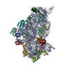
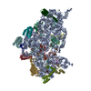
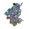
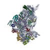
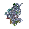
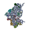
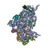
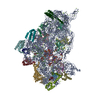
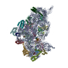
- EMDB-6710: Structure of the 30S small subunit of chloroplast ribosome from s... -
+ Open data

- Basic information
| Entry | Database: EMDB / ID: EMD-6710 | |||||||||
|---|---|---|---|---|---|---|---|---|---|---|
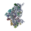
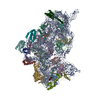
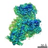
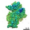
| Title | Structure of the 30S small subunit of chloroplast ribosome from spinach | |||||||||
 Map data Map data | ||||||||||
 Sample Sample |
| |||||||||
 Keywords Keywords | Cryo-EM / ribosome / chloroplast ribosome | |||||||||
| Function / homology |  Function and homology information Function and homology informationchloroplast rRNA processing / plastid small ribosomal subunit / plastid translation / negative regulation of translational elongation / mitochondrial small ribosomal subunit / mitochondrial translation / plastid / chloroplast stroma / chloroplast thylakoid membrane / ribosomal small subunit binding ...chloroplast rRNA processing / plastid small ribosomal subunit / plastid translation / negative regulation of translational elongation / mitochondrial small ribosomal subunit / mitochondrial translation / plastid / chloroplast stroma / chloroplast thylakoid membrane / ribosomal small subunit binding / chloroplast / ribosomal small subunit biogenesis / ribosomal small subunit assembly / small ribosomal subunit / small ribosomal subunit rRNA binding / cytosolic small ribosomal subunit / rRNA binding / structural constituent of ribosome / ribosome / translation / ribonucleoprotein complex / response to antibiotic / mRNA binding / mitochondrion / RNA binding Similarity search - Function | |||||||||
| Biological species |  Spinacia oleracea (spinach) Spinacia oleracea (spinach) | |||||||||
| Method | single particle reconstruction / cryo EM / Resolution: 3.7 Å | |||||||||
 Authors Authors | Ahmed T / Shi J | |||||||||
| Funding support |  Singapore, 1 items Singapore, 1 items
| |||||||||
 Citation Citation | Journal: Nucleic Acids Res. / Year: 2017 Title: Unique localization of the plastid-specific ribosomal proteins in the chloroplast ribosome small subunit provides mechanistic insights into the chloroplastic translation Authors: Ahmed T / Shi J / Bhushan S | |||||||||
| History |
|
- Structure visualization
| Movie |
Movie viewer |
|---|---|
| Structure viewer | EM map: SurfViewMolmilJmol/JSmol |
- Downloads & links
-EMDB archive
| Map data | emd_6710.map.gz | 9.8 MB | EMDB map data format | |
|---|---|---|---|---|
| Header (meta data) | emd-6710-v30.xmlemd-6710.xml | 38.2 KB 38.2 KB | Display Display | EMDB header |
| Images |  emd_6710.png emd_6710.png | 81 KB | ||
| Filedesc metadata | emd-6710.cif.gz | 9.4 KB | ||
| Archive directory |  http://ftp.pdbj.org/pub/emdb/structures/EMD-6710ftp://ftp.pdbj.org/pub/emdb/structures/EMD-6710 http://ftp.pdbj.org/pub/emdb/structures/EMD-6710ftp://ftp.pdbj.org/pub/emdb/structures/EMD-6710 | HTTPS FTP |
-Validation report
| Summary document | emd_6710_validation.pdf.gz | 389.5 KB | Display | EMDB validaton report |
|---|---|---|---|---|
| Full document | emd_6710_full_validation.pdf.gz | 389 KB | Display | |
| Data in XML | emd_6710_validation.xml.gz | 7 KB | Display | |
| Data in CIF | emd_6710_validation.cif.gz | 8 KB | Display | |
| Arichive directory | https://ftp.pdbj.org/pub/emdb/validation_reports/EMD-6710ftp://ftp.pdbj.org/pub/emdb/validation_reports/EMD-6710 | HTTPS FTP |
-Related structure data
| Related structure data | 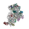 5x8rMC 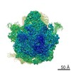 6709C 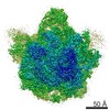 6711C 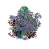 5x8pC  5x8tC M: atomic model generated by this map C: citing same article ( |
|---|---|
| Similar structure data |


-Links
| EMDB pages | EMDB (EBI/PDBe) / EMDataResource |
|---|---|
| Related items in Molecule of the Month |


-Map
| File | Download / File: emd_6710.map.gz / Format: CCP4 / Size: 216 MB / Type: IMAGE STORED AS FLOATING POINT NUMBER (4 BYTES) | ||||||||||||||||||||||||||||||||||||||||||||||||||||||||||||
|---|---|---|---|---|---|---|---|---|---|---|---|---|---|---|---|---|---|---|---|---|---|---|---|---|---|---|---|---|---|---|---|---|---|---|---|---|---|---|---|---|---|---|---|---|---|---|---|---|---|---|---|---|---|---|---|---|---|---|---|---|---|
| Voxel size | X=Y=Z: 1.05 Å | ||||||||||||||||||||||||||||||||||||||||||||||||||||||||||||
| Density |
| ||||||||||||||||||||||||||||||||||||||||||||||||||||||||||||
| Symmetry | Space group: 1 | ||||||||||||||||||||||||||||||||||||||||||||||||||||||||||||
| Details | EMDB XML:
CCP4 map header:
| ||||||||||||||||||||||||||||||||||||||||||||||||||||||||||||
-Supplemental data
- Sample components
+Entire : 30S small subunit of chloroplast ribosome from spinach
+Supramolecule #1: 30S small subunit of chloroplast ribosome from spinach
+Macromolecule #1: 30S ribosomal protein S2, chloroplastic
+Macromolecule #2: 30S ribosomal protein S3, chloroplastic
+Macromolecule #3: 30S ribosomal protein S5, chloroplastic
+Macromolecule #4: 30S ribosomal protein S6 alpha, chloroplastic
+Macromolecule #5: 30S ribosomal protein S7, chloroplastic
+Macromolecule #6: 30S ribosomal protein S8, chloroplastic
+Macromolecule #7: 30S ribosomal protein S9, chloroplastic
+Macromolecule #8: protein S10
+Macromolecule #9: 30S ribosomal protein S11, chloroplastic
+Macromolecule #10: 30S ribosomal protein S12, chloroplastic
+Macromolecule #11: 30S ribosomal protein S13, chloroplastic
+Macromolecule #12: 30S ribosomal protein S15, chloroplastic
+Macromolecule #13: 30S ribosomal protein S16, chloroplastic
+Macromolecule #14: protein S17
+Macromolecule #15: 30S ribosomal protein S18, chloroplastic
+Macromolecule #16: 30S ribosomal protein S19 alpha, chloroplastic
+Macromolecule #17: protein S20
+Macromolecule #18: protein S21
+Macromolecule #19: protein plastid pY
+Macromolecule #21: protein cS23
+Macromolecule #22: 30S ribosomal protein S4, chloroplastic
+Macromolecule #23: protein cS22
+Macromolecule #24: 30S ribosomal protein S14, chloroplastic
+Macromolecule #25: protein bTHXc
+Macromolecule #26: 30S ribosomal protein S1, chloroplastic
+Macromolecule #20: 16S rRNA
-Experimental details
-Structure determination
| Method | cryo EM |
|---|---|
 Processing Processing | single particle reconstruction |
| Aggregation state | particle |
-Sample preparation
| Buffer | pH: 7.6 Details: 20 mM Tris HCl, pH 7.6, 100 mM KCl, 10 mM MgOAc, 100 mM sucrose, 7 mM 2-mercaptoethanol, 1 unit/ml RNase inhibitor, 0.1% protease inhibitor |
|---|---|
| Grid | Model: Quantifoil / Material: COPPER / Mesh: 300 / Support film - Material: CARBON / Support film - topology: CONTINUOUS / Support film - Film thickness: 2 / Pretreatment - Type: GLOW DISCHARGE / Pretreatment - Time: 30 sec. |
| Vitrification | Cryogen name: ETHANE / Chamber humidity: 100 % / Chamber temperature: 277 K / Instrument: FEI VITROBOT MARK IV |
- Electron microscopy
| Microscope | FEI TITAN KRIOS |
|---|---|
| Image recording | Film or detector model: FEI FALCON II (4k x 4k) / Detector mode: INTEGRATING / Digitization - Dimensions - Width: 4096 pixel / Digitization - Dimensions - Height: 4096 pixel / Digitization - Frames/image: 1-25 / Number grids imaged: 1 / Number real images: 3161 / Average electron dose: 1.5 e/Å2 |
| Electron beam | Acceleration voltage: 300 kV / Electron source:  FIELD EMISSION GUN FIELD EMISSION GUN |
| Electron optics | C2 aperture diameter: 100.0 µm / Calibrated defocus max: 3.7 µm / Calibrated defocus min: 0.4 µm / Calibrated magnification: 133333 / Illumination mode: SPOT SCAN / Imaging mode: BRIGHT FIELD / Cs: 2.7 mm / Nominal defocus max: 3.7 µm / Nominal defocus min: 0.4 µm / Nominal magnification: 75000 |
| Sample stage | Specimen holder model: FEI TITAN KRIOS AUTOGRID HOLDER / Cooling holder cryogen: NITROGEN |
| Experimental equipment |  Model: Titan Krios / Image courtesy: FEI Company |
