 Movie
Movie Controller


+ Open data

- Basic information
| Entry | Database: EMDB / ID: EMD-6463 | |||||||||
|---|---|---|---|---|---|---|---|---|---|---|
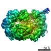
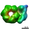
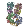
| Title | The Architecture of a Eukaryotic Replisome | |||||||||
 Map data Map data | Yeast CMG complex | |||||||||
 Sample Sample |
| |||||||||
 Keywords Keywords | Eukaryotic replisome / CMG / Electron microscopy | |||||||||
| Biological species |  | |||||||||
| Method | single particle reconstruction / negative staining / Resolution: 18.0 Å | |||||||||
 Authors Authors | Sun J / Shi Y / Georgescu RE / Yuan Z / Chait B / Li H / O'Donnell ME | |||||||||
 Citation Citation | Journal: Nat Struct Mol Biol / Year: 2015 Title: The architecture of a eukaryotic replisome. Authors: Jingchuan Sun / Yi Shi / Roxana E Georgescu / Zuanning Yuan / Brian T Chait / Huilin Li / Michael E O'Donnell /  Abstract: At the eukaryotic DNA replication fork, it is widely believed that the Cdc45-Mcm2-7-GINS (CMG) helicase is positioned in front to unwind DNA and that DNA polymerases trail behind the helicase. Here ...At the eukaryotic DNA replication fork, it is widely believed that the Cdc45-Mcm2-7-GINS (CMG) helicase is positioned in front to unwind DNA and that DNA polymerases trail behind the helicase. Here we used single-particle EM to directly image a Saccharomyces cerevisiae replisome. Contrary to expectations, the leading strand Pol ɛ is positioned ahead of CMG helicase, whereas Ctf4 and the lagging-strand polymerase (Pol) α-primase are behind the helicase. This unexpected architecture indicates that the leading-strand DNA travels a long distance before reaching Pol ɛ, first threading through the Mcm2-7 ring and then making a U-turn at the bottom and reaching Pol ɛ at the top of CMG. Our work reveals an unexpected configuration of the eukaryotic replisome, suggests possible reasons for this architecture and provides a basis for further structural and biochemical replisome studies. | |||||||||
| History |
|
- Structure visualization
| Movie |
 Movie viewer Movie viewer |
|---|---|
| Structure viewer | EM map: SurfViewMolmilJmol/JSmol |
| Supplemental images |

- Downloads & links
-EMDB archive
| Map data | emd_6463.map.gz | 934.5 KB | EMDB map data format | |
|---|---|---|---|---|
| Header (meta data) | emd-6463-v30.xmlemd-6463.xml | 8.5 KB 8.5 KB | Display Display | EMDB header |
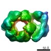
| Images | 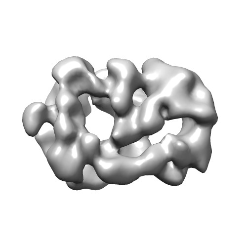 emd_6463.jpg emd_6463.jpg | 24.6 KB | ||
| Archive directory |  http://ftp.pdbj.org/pub/emdb/structures/EMD-6463ftp://ftp.pdbj.org/pub/emdb/structures/EMD-6463 http://ftp.pdbj.org/pub/emdb/structures/EMD-6463ftp://ftp.pdbj.org/pub/emdb/structures/EMD-6463 | HTTPS FTP |
-Related structure data

-Links
| EMDB pages | EMDB (EBI/PDBe) / EMDataResource |
|---|
-Map
| File | Download / File: emd_6463.map.gz / Format: CCP4 / Size: 1001 KB / Type: IMAGE STORED AS FLOATING POINT NUMBER (4 BYTES) | ||||||||||||||||||||||||||||||||||||||||||||||||||||||||||||||||||||
|---|---|---|---|---|---|---|---|---|---|---|---|---|---|---|---|---|---|---|---|---|---|---|---|---|---|---|---|---|---|---|---|---|---|---|---|---|---|---|---|---|---|---|---|---|---|---|---|---|---|---|---|---|---|---|---|---|---|---|---|---|---|---|---|---|---|---|---|---|---|
| Annotation | Yeast CMG complex | ||||||||||||||||||||||||||||||||||||||||||||||||||||||||||||||||||||
| Projections & slices | Image control
Images are generated by Spider. | ||||||||||||||||||||||||||||||||||||||||||||||||||||||||||||||||||||
| Voxel size | X=Y=Z: 4.24 Å | ||||||||||||||||||||||||||||||||||||||||||||||||||||||||||||||||||||
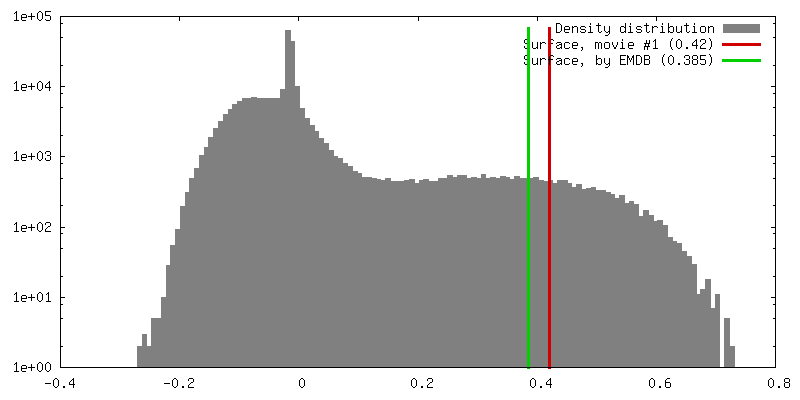
| Density |
| ||||||||||||||||||||||||||||||||||||||||||||||||||||||||||||||||||||
| Symmetry | Space group: 1 | ||||||||||||||||||||||||||||||||||||||||||||||||||||||||||||||||||||
| Details | EMDB XML:
CCP4 map header:
| ||||||||||||||||||||||||||||||||||||||||||||||||||||||||||||||||||||
 Z (Sec.)
 Y (Row.)
 X (Col.)


-Supplemental data
- Sample components
-Entire : Saccharomyces cerevisiae CMG complex
| Entire | Name: Saccharomyces cerevisiae CMG complex |
|---|---|
| Components |
|
-Supramolecule #1000: Saccharomyces cerevisiae CMG complex
| Supramolecule | Name: Saccharomyces cerevisiae CMG complex / type: sample / ID: 1000 / Number unique components: 1 |
|---|---|
| Molecular weight | Theoretical: 700 KDa |
-Macromolecule #1: CMG complex
| Macromolecule | Name: CMG complex / type: protein_or_peptide / ID: 1 / Number of copies: 1 / Recombinant expression: Yes |
|---|---|
| Source (natural) | Organism: |
| Molecular weight | Theoretical: 700 KDa |
| Recombinant expression | Organism: |
-Experimental details
-Structure determination
| Method | negative staining |
|---|---|
 Processing Processing | single particle reconstruction |
| Aggregation state | particle |
-Sample preparation
| Concentration | 0.5 mg/mL |
|---|---|
| Buffer | Details: 20 mM Tris-acetate, 40 mM potassium glutamate, 2 mM DTT, 0.1 mM EDTA |
| Staining | Type: NEGATIVE Details: Grids were stained with 1% w/v uranyl acetate for 1 minute. |
| Vitrification | Cryogen name: NONE / Instrument: OTHER |
- Electron microscopy
| Microscope | JEOL 2010F |
|---|---|
| Date | Oct 9, 2014 |
| Image recording | Category: CCD / Film or detector model: GATAN ULTRASCAN 4000 (4k x 4k) / Number real images: 300 / Average electron dose: 20 e/Å2 |
| Electron beam | Acceleration voltage: 200 kV / Electron source:  FIELD EMISSION GUN FIELD EMISSION GUN |
| Electron optics | Illumination mode: FLOOD BEAM / Imaging mode: BRIGHT FIELD / Nominal magnification: 50000 |
| Sample stage | Specimen holder model: GATAN LIQUID NITROGEN |
-Image processing
| Details | Particles were selected in EMAN 2.1 using SWAM. |
|---|---|
| CTF correction | Details: CTFFIND3 |
| Final reconstruction | Resolution.type: BY AUTHOR / Resolution: 18.0 Å / Resolution method: OTHER / Software - Name: Relion_1.3 / Number images used: 8781 |
