- EMDB-2772: CMG helicase bound to DNA and ATPgS -
+
Open data
ID or keywords:
Loading...
-
Basic information
Entry
Database: EMDB / ID: EMD-2772
Title
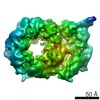
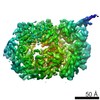
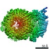
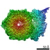
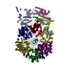
CMG helicase bound to DNA and ATPgS
Map data
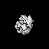
Reconstruction of the recombinant Drosophila CMG helicase bound to ATPgS and DNA
Sample
Sample: Drosophila melanogaster Cdc45-Mcm2-7-GINS complex bound to ATPgS and DNA
Protein or peptide: x 11 types
DNA: x 2 types
Keywords
Helicase / AAA+ ATPase / DNA replication
Function / homology
Function and homology information
Unwinding of DNA / Switching of origins to a post-replicative state / Assembly of the pre-replicative complex / DNA endoreduplication / Activation of ATR in response to replication stress / Activation of the pre-replicative complex / eggshell chorion gene amplification / Orc1 removal from chromatin / DNA amplification / DNA strand elongation involved in mitotic DNA replication ...Unwinding of DNA / Switching of origins to a post-replicative state / Assembly of the pre-replicative complex / DNA endoreduplication / Activation of ATR in response to replication stress / Activation of the pre-replicative complex / eggshell chorion gene amplification / Orc1 removal from chromatin / DNA amplification / DNA strand elongation involved in mitotic DNA replication / GINS complex / mitotic DNA replication preinitiation complex assembly / premeiotic DNA replication / resolution of meiotic recombination intermediates / mitotic DNA replication / CMG complex / DNA replication preinitiation complex / double-strand break repair via break-induced replication / MCM complex / mitotic DNA replication initiation / chromosome condensation / DNA strand elongation involved in DNA replication / DNA replication origin binding / DNA replication initiation / regulation of DNA-templated transcription elongation / mitotic spindle organization / DNA helicase activity / meiotic cell cycle / mitotic cell cycle / single-stranded DNA binding / DNA helicase / DNA replication / cell division / chromatin binding / ATP hydrolysis activity / zinc ion binding / ATP binding / nucleus / cytoplasm / cytosol Similarity search - Function
: / DNA replication licensing factor MCM2-like, winged-helix domain / : / PSF2 N-terminal domain / : / : / PSF3 N-terminal domain / PSF1 C-terminal domain / DNA replication complex GINS protein Psf2 / DNA replication complex GINS protein Psf2 ...: / DNA replication licensing factor MCM2-like, winged-helix domain / : / PSF2 N-terminal domain / : / : / PSF3 N-terminal domain / PSF1 C-terminal domain / DNA replication complex GINS protein Psf2 / DNA replication complex GINS protein Psf2 / CDC45 family / CDC45 family / CDC45 / GINS complex, subunit Psf3 / GINS complex, subunit Psf3 / DNA replication complex GINS protein SLD5, C-terminal / GINS complex, subunit Psf3 superfamily / GINS complex protein Sld5, alpha-helical domain / DNA replication complex GINS protein SLD5 C-terminus / GINS complex subunit Sld5 / GINS complex subunit Sld5 / GINS subunit, domain A / GINS subunit, domain A / GINS complex protein helical bundle domain / GINS complex, subunit Psf1 / GINS, helical bundle-like domain superfamily / : / MCM5, C-terminal domain / DNA replication licensing factor MCM7, winged helix / DNA replication licensing factor Mcm5 / DNA replication licensing factor Mcm5 / MCM4, winged helix domain / DNA replication licensing factor Mcm3 / Mini-chromosome maintenance complex protein 4 / DNA replication licensing factor Mcm3 / Mini-chromosome maintenance complex protein 4 / : / MCM3-like, winged helix domain / DNA replication licensing factor Mcm6 / DNA replication licensing factor Mcm7 / DNA replication licensing factor Mcm6 / DNA replication licensing factor Mcm7 / Mcm6, C-terminal winged-helix domain / MCM6 C-terminal winged-helix domain / DNA replication licensing factor Mcm2 / DNA replication licensing factor Mcm2 / Mini-chromosome maintenance protein 2 / Mini-chromosome maintenance, conserved site / MCM family signature. / MCM N-terminal domain / MCM N-terminal domain / MCM OB domain / MCM OB domain / Mini-chromosome maintenance protein / MCM, AAA-lid domain / MCM P-loop domain / MCM AAA-lid domain / MCM family C-terminal AAA(+) ATPase domain (MCM-CTD) profile. / minichromosome maintenance proteins / MCM domain / Winged helix-like DNA-binding domain superfamily / Nucleic acid-binding, OB-fold / ATPases associated with a variety of cellular activities / AAA+ ATPase domain / P-loop containing nucleoside triphosphate hydrolase Similarity search - Domain/homology
CDC45L / DNA replication licensing factor Mcm2 / DNA replication licensing factor MCM4 / DNA replication licensing factor Mcm6 / DNA replication complex GINS protein SLD5 / DNA replication licensing factor Mcm5 / Probable DNA replication complex GINS protein PSF2 / DNA replication complex GINS protein PSF1 / DNA replication complex GINS protein PSF3 / DNA replication licensing factor Mcm7 / DNA replication licensing factor Mcm3 Similarity search - Component
Journal: Elife / Year: 2014 Title: DNA binding polarity, dimerization, and ATPase ring remodeling in the CMG helicase of the eukaryotic replisome. Authors: Alessandro Costa / Ludovic Renault / Paolo Swuec / Tatjana Petojevic / James J Pesavento / Ivar Ilves / Kirsty MacLellan-Gibson / Roland A Fleck / Michael R Botchan / James M Berger / Abstract: The Cdc45/Mcm2-7/GINS (CMG) helicase separates DNA strands during replication in eukaryotes. How the CMG is assembled and engages DNA substrates remains unclear. Using electron microscopy, we have ...The Cdc45/Mcm2-7/GINS (CMG) helicase separates DNA strands during replication in eukaryotes. How the CMG is assembled and engages DNA substrates remains unclear. Using electron microscopy, we have determined the structure of the CMG in the presence of ATPγS and a DNA duplex bearing a 3' single-stranded tail. The structure shows that the MCM subunits of the CMG bind preferentially to single-stranded DNA, establishes the polarity by which DNA enters into the Mcm2-7 pore, and explains how Cdc45 helps prevent DNA from dissociating from the helicase. The Mcm2-7 subcomplex forms a cracked-ring, right-handed spiral when DNA and nucleotide are bound, revealing unexpected congruencies between the CMG and both bacterial DnaB helicases and the AAA+ motor of the eukaryotic proteasome. The existence of a subpopulation of dimeric CMGs establishes the subunit register of Mcm2-7 double hexamers and together with the spiral form highlights how Mcm2-7 transitions through different conformational and assembly states as it matures into a functional helicase.
History
Deposition
Aug 28, 2014
-
Header (metadata) release
Sep 10, 2014
-
Map release
Sep 10, 2014
-
Update
Sep 10, 2014
-
Current status
Sep 10, 2014
Processing site: PDBe / Status: Released
-
Structure visualization
Movie
Surface view with section colored by density value
Entire : Drosophila melanogaster Cdc45-Mcm2-7-GINS complex bound to ATPgS ...
Entire
Name: Drosophila melanogaster Cdc45-Mcm2-7-GINS complex bound to ATPgS and DNA
Components
Sample: Drosophila melanogaster Cdc45-Mcm2-7-GINS complex bound to ATPgS and DNA
Protein or peptide: Mcm2
Protein or peptide: Mcm3
Protein or peptide: Mcm4
Protein or peptide: Mcm5
Protein or peptide: Mcm6
Protein or peptide: Mcm7
Protein or peptide: Cdc45
Protein or peptide: Psf1
Protein or peptide: Psf2
Protein or peptide: Psf3
Protein or peptide: Sld5
DNA: LEAD60
DNA: 3BTNLAG20
+
Supramolecule #1000: Drosophila melanogaster Cdc45-Mcm2-7-GINS complex bound to ATPgS ...
Supramolecule
Name: Drosophila melanogaster Cdc45-Mcm2-7-GINS complex bound to ATPgS and DNA type: sample / ID: 1000 / Details: The sample was monodisperse / Oligomeric state: 13-mer / Number unique components: 13
UniProtKB: DNA replication complex GINS protein SLD5 / InterPro: GINS complex subunit Sld5
+
Macromolecule #12: LEAD60
Macromolecule
Name: LEAD60 / type: dna / ID: 12 Details: The two oligonucleotides have been annealed to obtain a 20mer duplex DNA with a 40mer 3' overhang. Classification: DNA / Structure: OTHER / Synthetic?: Yes
Name: 3BTNLAG20 / type: dna / ID: 13 Details: The two oligonucleotides have been annealed to obtain a 20mer duplex DNA with a 40mer 3' overhang. 3BTNLAG20 contains a biotin on the 3' end (duplex end). Classification: DNA / Structure: OTHER / Synthetic?: Yes
Source (natural)
Organism: unidentified (others)
Molecular weight
Theoretical: 6.569 KDa
Sequence
String:
GGTTGGCCGA TCAAGTGCCC
-
Experimental details
-
Structure determination
Method
negative staining
Processing
single particle reconstruction
Aggregation state
particle
-
Sample preparation
Concentration
0.02 mg/mL
Buffer
pH: 7.6 Details: 25 mM HEPES , 50 mM sodium acetate, 10 mM magnesium acetate, 0.1 mM ATPgS, 1 mM DTT
Staining
Type: NEGATIVE Details: The grid with adsorbed protein was floated for 10 seconds on four consecutive 75 microliter drops of 2% w/v uranyl formate solution.
Grid
Details: 400 mesh copper grids with thin carbon support.
Vitrification
Cryogen name: NONE / Instrument: OTHER Details: The sample stained with a fresh 2% (wt/vol) uranyl formate solution.
-
Electron microscopy
Microscope
JEOL 2100
Alignment procedure
Legacy - Astigmatism: Objective lens astigmatism was corrected at 60,000 times magnification
Date
May 8, 2013
Image recording
Category: CCD / Film or detector model: GATAN ULTRASCAN 4000 (4k x 4k) / Digitization - Sampling interval: 15 µm / Number real images: 579 / Average electron dose: 35 e/Å2 / Bits/pixel: 32
Electron beam
Acceleration voltage: 200 kV / Electron source: LAB6
In the structure databanks used in Yorodumi, some data are registered as the other names, "COVID-19 virus" and "2019-nCoV". Here are the details of the virus and the list of structure data.
Jan 31, 2019. EMDB accession codes are about to change! (news from PDBe EMDB page)
EMDB accession codes are about to change! (news from PDBe EMDB page)
The allocation of 4 digits for EMDB accession codes will soon come to an end. Whilst these codes will remain in use, new EMDB accession codes will include an additional digit and will expand incrementally as the available range of codes is exhausted. The current 4-digit format prefixed with “EMD-” (i.e. EMD-XXXX) will advance to a 5-digit format (i.e. EMD-XXXXX), and so on. It is currently estimated that the 4-digit codes will be depleted around Spring 2019, at which point the 5-digit format will come into force.
The EM Navigator/Yorodumi systems omit the EMD- prefix.
Related info.:Q: What is EMD? / ID/Accession-code notation in Yorodumi/EM Navigator
Yorodumi is a browser for structure data from EMDB, PDB, SASBDB, etc.
This page is also the successor to EM Navigator detail page, and also detail information page/front-end page for Omokage search.
The word "yorodu" (or yorozu) is an old Japanese word meaning "ten thousand". "mi" (miru) is to see.
Related info.:EMDB / PDB / SASBDB / Comparison of 3 databanks / Yorodumi Search / Aug 31, 2016. New EM Navigator & Yorodumi / Yorodumi Papers / Jmol/JSmol / Function and homology information / Changes in new EM Navigator and Yorodumi
 Movie
Movie Controller


 Open data
 Basic information
 Map data
 Sample
 Keywords
 Function and homology information
 Authors
 Citation


 Structure visualization
 UCSF Chimera
 Downloads & links
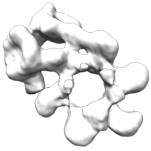 EMD-2772-deposition_image.png
 http://ftp.pdbj.org/pub/emdb/structures/EMD-2772

 Z (Sec.)
 Y (Row.)
 X (Col.)


 Sample components
 unidentified baculovirus
 Processing
 Electron microscopy