 ムービー
ムービー コントローラー


+ データを開く

- 基本情報
基本情報
| 登録情報 | データベース: EMDB / ID: EMD-6429 | |||||||||
|---|---|---|---|---|---|---|---|---|---|---|
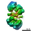
| タイトル | Architecture of the complex formed by large and small Terminase subunits from Bacteriophage P22 | |||||||||
 マップデータ マップデータ | Asymmetric reconstruction of terminase holoenzyme from Bacteriophage P22 | |||||||||
 試料 試料 |
| |||||||||
 キーワード キーワード | viral genome-packaging motor / Terminase complex / Salmonella virus / P22 Bacteriophage | |||||||||
| 生物種 |  Enterobacteria phage P22 (ファージ) Enterobacteria phage P22 (ファージ) | |||||||||
| 手法 | 単粒子再構成法 / ネガティブ染色法 / 解像度: 30.0 Å | |||||||||
 データ登録者 データ登録者 | McNulty R / Johnson JE | |||||||||
 引用 引用 | ジャーナル: J Mol Biol / 年: 2015 タイトル: Architecture of the Complex Formed by Large and Small Terminase Subunits from Bacteriophage P22. 著者: Reginald McNulty / Ravi Kumar Lokareddy / Ankoor Roy / Yang Yang / Gabriel C Lander / Albert J R Heck / John E Johnson / Gino Cingolani /   要旨: Packaging of viral genomes inside empty procapsids is driven by a powerful ATP-hydrolyzing motor, formed in many double-stranded DNA viruses by a complex of a small terminase (S-terminase) subunit ...Packaging of viral genomes inside empty procapsids is driven by a powerful ATP-hydrolyzing motor, formed in many double-stranded DNA viruses by a complex of a small terminase (S-terminase) subunit and a large terminase (L-terminase) subunit, transiently docked at the portal vertex during genome packaging. Despite recent progress in elucidating the structure of individual terminase subunits and their domains, little is known about the architecture of an assembled terminase complex. Here, we describe a bacterial co-expression system that yields milligram quantities of the S-terminase:L-terminase complex of the Salmonella phage P22. In vivo assembled terminase complex was affinity-purified and stabilized by addition of non-hydrolyzable ATP, which binds specifically to the ATPase domain of L-terminase. Mapping studies revealed that the N-terminus of L-terminase ATPase domain (residues 1-58) contains a minimal S-terminase binding domain sufficient for stoichiometric association with residues 140-162 of S-terminase, the L-terminase binding domain. Hydrodynamic analysis by analytical ultracentrifugation sedimentation velocity and native mass spectrometry revealed that the purified terminase complex consists predominantly of one copy of the nonameric S-terminase bound to two equivalents of L-terminase (1S-terminase:2L-terminase). Direct visualization of this molecular assembly in negative-stained micrographs yielded a three-dimensional asymmetric reconstruction that resembles a "nutcracker" with two L-terminase protomers projecting from the C-termini of an S-terminase ring. This is the first direct visualization of a purified viral terminase complex analyzed in the absence of DNA and procapsid. | |||||||||
| 履歴 |
|
- 構造の表示
構造の表示
| ムービー |
 ムービービューア ムービービューア |
|---|---|
| 構造ビューア | EMマップ: SurfViewMolmilJmol/JSmol |
| 添付画像 |

- ダウンロードとリンク
-EMDBアーカイブ
| マップデータ | emd_6429.map.gz | 1 MB | EMDBマップデータ形式 | |
|---|---|---|---|---|
| ヘッダ (付随情報) | emd-6429-v30.xmlemd-6429.xml | 12.6 KB 12.6 KB | 表示 表示 | EMDBヘッダ |
| FSC (解像度算出) | emd_6429_fsc.xml | 2.6 KB | 表示 | FSCデータファイル |
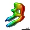
| 画像 | 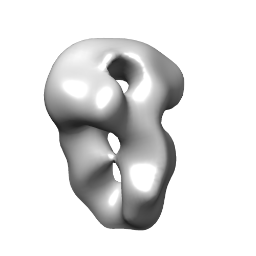 emd_6429.png emd_6429.png | 38.9 KB | ||
| マスクデータ | emd_6429_msk_1.map | 1.4 MB | マスクマップ | |
| アーカイブディレクトリ |  http://ftp.pdbj.org/pub/emdb/structures/EMD-6429ftp://ftp.pdbj.org/pub/emdb/structures/EMD-6429 http://ftp.pdbj.org/pub/emdb/structures/EMD-6429ftp://ftp.pdbj.org/pub/emdb/structures/EMD-6429 | HTTPS FTP |
-検証レポート
| 文書・要旨 | emd_6429_validation.pdf.gz | 77.5 KB | 表示 | EMDB検証レポート |
|---|---|---|---|---|
| 文書・詳細版 | emd_6429_full_validation.pdf.gz | 76.6 KB | 表示 | |
| XML形式データ | emd_6429_validation.xml.gz | 495 B | 表示 | |
| アーカイブディレクトリ | https://ftp.pdbj.org/pub/emdb/validation_reports/EMD-6429ftp://ftp.pdbj.org/pub/emdb/validation_reports/EMD-6429 | HTTPS FTP |
-関連構造データ


-リンク
| EMDBのページ | EMDB (EBI/PDBe) / EMDataResource |
|---|
-マップ
| ファイル | ダウンロード / ファイル: emd_6429.map.gz / 形式: CCP4 / 大きさ: 1.4 MB / タイプ: IMAGE STORED AS FLOATING POINT NUMBER (4 BYTES) | ||||||||||||||||||||||||||||||||||||||||||||||||||||||||||||||||||||
|---|---|---|---|---|---|---|---|---|---|---|---|---|---|---|---|---|---|---|---|---|---|---|---|---|---|---|---|---|---|---|---|---|---|---|---|---|---|---|---|---|---|---|---|---|---|---|---|---|---|---|---|---|---|---|---|---|---|---|---|---|---|---|---|---|---|---|---|---|---|
| 注釈 | Asymmetric reconstruction of terminase holoenzyme from Bacteriophage P22 | ||||||||||||||||||||||||||||||||||||||||||||||||||||||||||||||||||||
| 投影像・断面図 | 画像のコントロール
画像は Spider により作成 | ||||||||||||||||||||||||||||||||||||||||||||||||||||||||||||||||||||
| ボクセルのサイズ | X=Y=Z: 4.1 Å | ||||||||||||||||||||||||||||||||||||||||||||||||||||||||||||||||||||
| 密度 |
| ||||||||||||||||||||||||||||||||||||||||||||||||||||||||||||||||||||
| 対称性 | 空間群: 1 | ||||||||||||||||||||||||||||||||||||||||||||||||||||||||||||||||||||
| 詳細 | EMDB XML:
CCP4マップ ヘッダ情報:
| ||||||||||||||||||||||||||||||||||||||||||||||||||||||||||||||||||||
 Z (Sec.)
 Y (Row.)
 X (Col.)


-添付データ
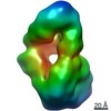
-セグメンテーションマップ: Mask of complex generated automatically with RELION
| 注釈 | Mask of complex generated automatically with RELION | ||||||||||||
|---|---|---|---|---|---|---|---|---|---|---|---|---|---|
| ファイル | emd_6429_msk_1.map | ||||||||||||
| 投影像・断面図 |
| ||||||||||||
| 密度ヒストグラム |


- 試料の構成要素
-全体 : Bacteriophage P22 terminase holoenzyme
| 全体 | 名称: Bacteriophage P22 terminase holoenzyme |
|---|---|
| 要素 |
|
-超分子 #1000: Bacteriophage P22 terminase holoenzyme
| 超分子 | 名称: Bacteriophage P22 terminase holoenzyme / タイプ: sample / ID: 1000 集合状態: One nonamer of S-terminase binds to two L-terminase subunits Number unique components: 2 |
|---|---|
| 分子量 | 実験値: 287 KDa / 理論値: 287 KDa / 手法: Native MS |
-分子 #1: small terminase subunit
| 分子 | 名称: small terminase subunit / タイプ: protein_or_peptide / ID: 1 / コピー数: 9 / 集合状態: 9 small subunits : 2 large subunits / 組換発現: Yes |
|---|---|
| 由来(天然) | 生物種: Enterobacteria phage P22 (ファージ) / 別称: P22 |
| 組換発現 | 生物種:  |
-分子 #2: large terminase subunit
| 分子 | 名称: large terminase subunit / タイプ: protein_or_peptide / ID: 2 / コピー数: 2 / 集合状態: 9 small subunits : 2 large subunits / 組換発現: Yes |
|---|---|
| 由来(天然) | 生物種: Enterobacteria phage P22 (ファージ) / 別称: P22 |
| 組換発現 | 生物種: |
-実験情報
-構造解析
| 手法 | ネガティブ染色法 |
|---|---|
 解析 解析 | 単粒子再構成法 |
| 試料の集合状態 | particle |
-試料調製
| 濃度 | 0.014 mg/mL |
|---|---|
| 緩衝液 | pH: 8 詳細: 20 mM Tris-HCl, 150 mM NaCl, 3 mm DTT, 5% glycerol, 1 mM MgCl2 |
| 染色 | タイプ: NEGATIVE 詳細: Protein was adsorbed to the grid for 1 minute, blotted, and passed through four consecutive 40 microliter drops of 2% uranyl formate. |
| グリッド | 詳細: 400 mesh copper grids charged with Gatan plasma cleaner |
| 凍結 | 凍結剤: NONE / 装置: OTHER |
- 電子顕微鏡法
| 顕微鏡 | FEI TECNAI 12 |
|---|---|
| 日付 | 2015年3月25日 |
| 撮影 | カテゴリ: CCD フィルム・検出器のモデル: TVIPS TEMCAM-F416 (4k x 4k) 実像数: 44 / 平均電子線量: 20 e/Å2 / 詳細: Images were acquired using Leginon. |
| 電子線 | 加速電圧: 120 kV / 電子線源:  FIELD EMISSION GUN FIELD EMISSION GUN |
| 電子光学系 | 照射モード: FLOOD BEAM / 撮影モード: BRIGHT FIELD / 最大 デフォーカス(公称値): 0.2 µm / 最小 デフォーカス(公称値): 0.1 µm / 倍率(公称値): 52000 |
| 試料ステージ | 試料ホルダーモデル: SIDE ENTRY, EUCENTRIC |
-画像解析
| 詳細 | Particles were selected and filtered using the Appion Suite. Refinement was done with RELION. |
|---|---|
| 最終 再構成 | アルゴリズム: OTHER / 解像度のタイプ: BY AUTHOR / 解像度: 30.0 Å / 解像度の算出法: OTHER / ソフトウェア - 名称: RELION / 使用した粒子像数: 2062 |
| FSC曲線 (解像度の算出) |  |
