 Movie
Movie Controller


+ Open data

- Basic information
| Entry | Database: PDB / ID: 5me3 | ||||||||||||
|---|---|---|---|---|---|---|---|---|---|---|---|---|---|
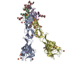
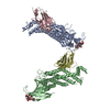
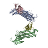
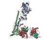
| Title | Structure of the Scc2 C-terminus | ||||||||||||
 Components Components |
| ||||||||||||
 Keywords Keywords | CELL CYCLE / cohesin loader / HEAT repeat / Scc2 | ||||||||||||
| Function / homology |  Function and homology information Function and homology informationScc2-Scc4 cohesin loading complex / cohesin loader activity / replication-born double-strand break repair via sister chromatid exchange / establishment of mitotic sister chromatid cohesion / establishment of protein localization to chromatin / chromatin looping / regulation of gene expression / chromatin binding Similarity search - Function | ||||||||||||
| Biological species |  Ashbya gossypii (fungus)Eremothecium gossypii ATCC 10895 (fungus) Ashbya gossypii (fungus)Eremothecium gossypii ATCC 10895 (fungus) | ||||||||||||
| Method |  X-RAY DIFFRACTION / SYNCHROTRON / MIR / Resolution: 2.85 Å X-RAY DIFFRACTION / SYNCHROTRON / MIR / Resolution: 2.85 Å | ||||||||||||
 Authors Authors | Chao, W.C.H. / Singleton, M.R. | ||||||||||||
| Funding support |  United Kingdom, 3items United Kingdom, 3items
| ||||||||||||
 Citation Citation | Journal: Nat Commun / Year: 2017 Title: Structure of the cohesin loader Scc2. Authors: Chao, W.C. / Murayama, Y. / Munoz, S. / Jones, A.W. / Wade, B.O. / Purkiss, A.G. / Hu, X.W. / Borg, A. / Snijders, A.P. / Uhlmann, F. / Singleton, M.R. | ||||||||||||
| History |
|
- Structure visualization
| Structure viewer | Molecule: MolmilJmol/JSmol |
|---|
- Downloads & links
-Download
| PDBx/mmCIF format | 5me3.cif.gz | 412.6 KB | Display | PDBx/mmCIF format |
|---|---|---|---|---|
| PDB format | pdb5me3.ent.gz | 325.3 KB | Display | PDB format |
| PDBx/mmJSON format | 5me3.json.gz | Tree view | PDBx/mmJSON format | |
| Others |  Other downloads Other downloads |
-Validation report
| Arichive directory | https://data.pdbj.org/pub/pdb/validation_reports/me/5me3ftp://data.pdbj.org/pub/pdb/validation_reports/me/5me3 | HTTPS FTP |
|---|
-Related structure data
| Similar structure data |
|---|


-Links
 PDBj
PDBj- Assembly
| Deposited unit | 
| ||||||||
|---|---|---|---|---|---|---|---|---|---|
| 1 | 
| ||||||||
| 2 | 
| ||||||||
| Unit cell |
|
-Components
| #1: Protein | Mass: 131560.844 Da / Num. of mol.: 2 / Fragment: UNP residues 378-1479 Source method: isolated from a genetically manipulated source Source: (gene. exp.) Ashbya gossypii (strain ATCC 10895 / CBS 109.51 / FGSC 9923 / NRRL Y-1056) (fungus)Gene: SCC2, AGL133W / Production host:   Spodoptera frugiperda (fall armyworm) / References: UniProt: Q750S2 Spodoptera frugiperda (fall armyworm) / References: UniProt: Q750S2#2: Protein/peptide | | Mass: 1464.797 Da / Num. of mol.: 1 Source method: isolated from a genetically manipulated source Details: unassigned sequence of Scc2 Source: (gene. exp.) Eremothecium gossypii ATCC 10895 (fungus)Production host: Spodoptera frugiperda (fall armyworm)#3: Protein/peptide | | Mass: 2315.846 Da / Num. of mol.: 1 Source method: isolated from a genetically manipulated source Details: Scc2 unassigned sequence Source: (gene. exp.) Eremothecium gossypii ATCC 10895 (fungus)Production host: Spodoptera frugiperda (fall armyworm)#4: Protein/peptide | Mass: 954.168 Da / Num. of mol.: 2 Source method: isolated from a genetically manipulated source Source: (gene. exp.) Eremothecium gossypii ATCC 10895 (fungus)Production host: Spodoptera frugiperda (fall armyworm) |
|---|
-Experimental details
-Experiment
| Experiment | Method: X-RAY DIFFRACTION / Number of used crystals: 1 |
|---|
- Sample preparation
| Crystal | Density Matthews: 2.71 Å3/Da / Density % sol: 54.58 % |
|---|---|
| Crystal grow | Temperature: 277 K / Method: vapor diffusion, hanging drop Details: 100 mM imidazole (pH 6.8), 200 mM lithium sulphate, and 4.5% polyethylene glycol 5000 MME |
-Data collection
| Diffraction | Mean temperature: 93 K |
|---|---|
| Diffraction source | Source: SYNCHROTRON / Site: Diamond / Beamline: I02 / Wavelength: 0.9795 Å |
| Detector | Type: DECTRIS PILATUS 6M-F / Detector: PIXEL / Date: Feb 14, 2015 |
| Radiation | Protocol: SINGLE WAVELENGTH / Monochromatic (M) / Laue (L): M / Scattering type: x-ray |
| Radiation wavelength | Wavelength: 0.9795 Å / Relative weight: 1 |
| Reflection | Resolution: 2.85→48.81 Å / Num. obs: 65853 / % possible obs: 99 % / Redundancy: 3.3 % / Rmerge(I) obs: 0.0715 / Net I/σ(I): 10.36 |
| Reflection shell | Resolution: 2.85→2.952 Å / Redundancy: 2.9 % / Rmerge(I) obs: 1.244 / Mean I/σ(I) obs: 0.75 / CC1/2: 0.312 / % possible all: 96 |
- Processing
| Software |
| |||||||||||||||||||||||||||||||||||||||||||||||||||||||||||||||||||||||||||||||||||||||||||||||||||||||||||||||||||||||||||||||||||||||||||||||||||||||||||||||||||||||||||||||
|---|---|---|---|---|---|---|---|---|---|---|---|---|---|---|---|---|---|---|---|---|---|---|---|---|---|---|---|---|---|---|---|---|---|---|---|---|---|---|---|---|---|---|---|---|---|---|---|---|---|---|---|---|---|---|---|---|---|---|---|---|---|---|---|---|---|---|---|---|---|---|---|---|---|---|---|---|---|---|---|---|---|---|---|---|---|---|---|---|---|---|---|---|---|---|---|---|---|---|---|---|---|---|---|---|---|---|---|---|---|---|---|---|---|---|---|---|---|---|---|---|---|---|---|---|---|---|---|---|---|---|---|---|---|---|---|---|---|---|---|---|---|---|---|---|---|---|---|---|---|---|---|---|---|---|---|---|---|---|---|---|---|---|---|---|---|---|---|---|---|---|---|---|---|---|---|---|
| Refinement | Method to determine structure: MIR / Resolution: 2.85→48.81 Å / SU ML: 0.54 / Cross valid method: FREE R-VALUE / σ(F): 1.36 / Phase error: 29.9 / Stereochemistry target values: ML
| |||||||||||||||||||||||||||||||||||||||||||||||||||||||||||||||||||||||||||||||||||||||||||||||||||||||||||||||||||||||||||||||||||||||||||||||||||||||||||||||||||||||||||||||
| Solvent computation | Shrinkage radii: 0.9 Å / VDW probe radii: 1.11 Å / Solvent model: FLAT BULK SOLVENT MODEL | |||||||||||||||||||||||||||||||||||||||||||||||||||||||||||||||||||||||||||||||||||||||||||||||||||||||||||||||||||||||||||||||||||||||||||||||||||||||||||||||||||||||||||||||
| Refinement step | Cycle: LAST / Resolution: 2.85→48.81 Å
| |||||||||||||||||||||||||||||||||||||||||||||||||||||||||||||||||||||||||||||||||||||||||||||||||||||||||||||||||||||||||||||||||||||||||||||||||||||||||||||||||||||||||||||||
| Refine LS restraints |
| |||||||||||||||||||||||||||||||||||||||||||||||||||||||||||||||||||||||||||||||||||||||||||||||||||||||||||||||||||||||||||||||||||||||||||||||||||||||||||||||||||||||||||||||
| LS refinement shell |
|
