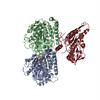
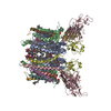
ジャーナル: EMBO J / 年: 2015 タイトル: X-ray and Cryo-EM structures reveal mutual conformational changes of Kinesin and GTP-state microtubules upon binding. 著者: Manatsu Morikawa / Hiroaki Yajima / Ryo Nitta / Shigeyuki Inoue / Toshihiko Ogura / Chikara Sato / Nobutaka Hirokawa / 要旨: The molecular motor kinesin moves along microtubules using energy from ATP hydrolysis in an initial step coupled with ADP release. In neurons, kinesin-1/KIF5C preferentially binds to the GTP-state ...The molecular motor kinesin moves along microtubules using energy from ATP hydrolysis in an initial step coupled with ADP release. In neurons, kinesin-1/KIF5C preferentially binds to the GTP-state microtubules over GDP-state microtubules to selectively enter an axon among many processes; however, because the atomic structure of nucleotide-free KIF5C is unavailable, its molecular mechanism remains unresolved. Here, the crystal structure of nucleotide-free KIF5C and the cryo-electron microscopic structure of nucleotide-free KIF5C complexed with the GTP-state microtubule are presented. The structures illustrate mutual conformational changes induced by interaction between the GTP-state microtubule and KIF5C. KIF5C acquires the 'rigor conformation', where mobile switches I and II are stabilized through L11 and the initial portion of the neck-linker, facilitating effective ADP release and the weak-to-strong transition of KIF5C microtubule affinity. Conformational changes to tubulin strengthen the longitudinal contacts of the GTP-state microtubule in a similar manner to GDP-taxol microtubules. These results and functional analyses provide the molecular mechanism of the preferential binding of KIF5C to GTP-state microtubules.
全体 : Cryo-Electron Microscopy of Nucleotide-free Kinesin motor domain ...
全体
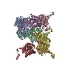
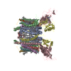
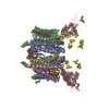
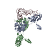
名称: Cryo-Electron Microscopy of Nucleotide-free Kinesin motor domain complexed with GMPCPP-microtubule
要素
試料: Cryo-Electron Microscopy of Nucleotide-free Kinesin motor domain complexed with GMPCPP-microtubule
タンパク質・ペプチド: TUBULIN ALPHA CHAIN
タンパク質・ペプチド: TUBULIN BETA CHAIN
タンパク質・ペプチド: Kinesin heavy chain isoform 5C motor domain
-
超分子 #1000: Cryo-Electron Microscopy of Nucleotide-free Kinesin motor domain ...
超分子
名称: Cryo-Electron Microscopy of Nucleotide-free Kinesin motor domain complexed with GMPCPP-microtubule タイプ: sample / ID: 1000 集合状態: One kineisn motor domain binds to one tubulin dimers Number unique components: 3
 ムービー
ムービー コントローラー


 データを開く
 基本情報
基本情報 マップデータ
 試料
試料 キーワード
キーワード 機能・相同性情報
 データ登録者
 引用
引用

 構造の表示
構造の表示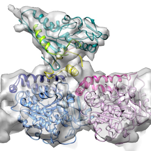
 ダウンロードとリンク
 http://ftp.pdbj.org/pub/emdb/structures/EMD-5916


 Z (Sec.)
 Y (Row.)
 X (Col.)


 試料の構成要素
 解析
解析 電子顕微鏡法
 FIELD EMISSION GUN