ムービー
ムービー コントローラー


+ データを開く

- 基本情報
基本情報
| 登録情報 | データベース: EMDB / ID: EMD-5404 | |||||||||
|---|---|---|---|---|---|---|---|---|---|---|
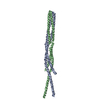
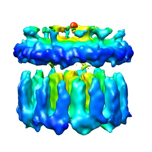
| タイトル | Molecular Architecture of Chemoreceptor Array in E. coli | |||||||||
 マップデータ マップデータ | This in situ structure of chemoreceptor array was determined by using cryo-electron tomography of E. coli minicell | |||||||||
 試料 試料 |
| |||||||||
 キーワード キーワード | Bacterial Chemotaxis Chemoreceptor array Signaling Transduction Minicell cryo-electron tomography | |||||||||
| 生物種 |  | |||||||||
| 手法 | サブトモグラム平均法 / クライオ電子顕微鏡法 / ネガティブ染色法 / 解像度: 32.0 Å | |||||||||
 データ登録者 データ登録者 | Liu J / Hu B / Morado DR / Jani S / Manson MD / Margolin W | |||||||||
 引用 引用 | ジャーナル: Proc Natl Acad Sci U S A / 年: 2012 タイトル: Molecular architecture of chemoreceptor arrays revealed by cryoelectron tomography of Escherichia coli minicells. 著者: Jun Liu / Bo Hu / Dustin R Morado / Sneha Jani / Michael D Manson / William Margolin /  要旨: The chemoreceptors of Escherichia coli localize to the cell poles and form a highly ordered array in concert with the CheA kinase and the CheW coupling factor. However, a high-resolution structure of ...The chemoreceptors of Escherichia coli localize to the cell poles and form a highly ordered array in concert with the CheA kinase and the CheW coupling factor. However, a high-resolution structure of the array has been lacking, and the molecular basis of array assembly has thus remained elusive. Here, we use cryoelectron tomography of flagellated E. coli minicells to derive a 3D map of the intact array. Docking of high-resolution structures into the 3D map provides a model of the core signaling complex, in which a CheA/CheW dimer bridges two adjacent receptor trimers via multiple hydrophobic interactions. A further, hitherto unknown, hydrophobic interaction between CheW and the homologous P5 domain of CheA in an adjacent core complex connects the complexes into an extended array. This architecture provides a structural basis for array formation and could explain the high sensitivity and cooperativity of chemotaxis signaling in E. coli. | |||||||||
| 履歴 |
|
- 構造の表示
構造の表示
| ムービー |
 ムービービューア ムービービューア |
|---|---|
| 構造ビューア | EMマップ: SurfViewMolmilJmol/JSmol |
| 添付画像 |
- ダウンロードとリンク
-EMDBアーカイブ
| マップデータ | emd_5404.map.gz | 3.2 MB | EMDBマップデータ形式 | |
|---|---|---|---|---|
| ヘッダ (付随情報) | emd-5404-v30.xmlemd-5404.xml | 10.6 KB 10.6 KB | 表示 表示 | EMDBヘッダ |
| 画像 |  emd_5404_1.jpg emd_5404_1.jpg | 51.4 KB | ||
| アーカイブディレクトリ |  http://ftp.pdbj.org/pub/emdb/structures/EMD-5404ftp://ftp.pdbj.org/pub/emdb/structures/EMD-5404 http://ftp.pdbj.org/pub/emdb/structures/EMD-5404ftp://ftp.pdbj.org/pub/emdb/structures/EMD-5404 | HTTPS FTP |
-関連構造データ
| 類似構造データ |
|---|

-リンク
| EMDBのページ | EMDB (EBI/PDBe) / EMDataResource |
|---|
-マップ
| ファイル | ダウンロード / ファイル: emd_5404.map.gz / 形式: CCP4 / 大きさ: 7.8 MB / タイプ: IMAGE STORED AS FLOATING POINT NUMBER (4 BYTES) | ||||||||||||||||||||||||||||||||||||||||||||||||||||||||||||||||||||
|---|---|---|---|---|---|---|---|---|---|---|---|---|---|---|---|---|---|---|---|---|---|---|---|---|---|---|---|---|---|---|---|---|---|---|---|---|---|---|---|---|---|---|---|---|---|---|---|---|---|---|---|---|---|---|---|---|---|---|---|---|---|---|---|---|---|---|---|---|---|
| 注釈 | This in situ structure of chemoreceptor array was determined by using cryo-electron tomography of E. coli minicell | ||||||||||||||||||||||||||||||||||||||||||||||||||||||||||||||||||||
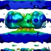
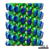
| 投影像・断面図 | 画像のコントロール
画像は Spider により作成 | ||||||||||||||||||||||||||||||||||||||||||||||||||||||||||||||||||||
| ボクセルのサイズ | X=Y=Z: 5.7 Å | ||||||||||||||||||||||||||||||||||||||||||||||||||||||||||||||||||||
| 密度 |
| ||||||||||||||||||||||||||||||||||||||||||||||||||||||||||||||||||||
| 対称性 | 空間群: 1 | ||||||||||||||||||||||||||||||||||||||||||||||||||||||||||||||||||||
| 詳細 | EMDB XML:
CCP4マップ ヘッダ情報:
| ||||||||||||||||||||||||||||||||||||||||||||||||||||||||||||||||||||
 Z (Sec.)
 Y (Row.)
 X (Col.)


-添付データ
- 試料の構成要素
-全体 : Molecular Architecture of Chemoreceptor Arrays
| 全体 | 名称: Molecular Architecture of Chemoreceptor Arrays |
|---|---|
| 要素 |
|
-超分子 #1000: Molecular Architecture of Chemoreceptor Arrays
| 超分子 | 名称: Molecular Architecture of Chemoreceptor Arrays / タイプ: sample / ID: 1000 / 詳細: The sample is E. coli minicell. / Number unique components: 3 |
|---|
-超分子 #1: Bacterial chemoreceptor arrays
| 超分子 | 名称: Bacterial chemoreceptor arrays / タイプ: organelle_or_cellular_component / ID: 1 / 組換発現: No / データベース: NCBI |
|---|---|
| 由来(天然) | 生物種: |
-実験情報
-構造解析
| 手法 | ネガティブ染色法, クライオ電子顕微鏡法 |
|---|---|
 解析 解析 | サブトモグラム平均法 |
| 試料の集合状態 | particle |
-試料調製
| 緩衝液 | 詳細: tryptone broth |
|---|---|
| 染色 | タイプ: NEGATIVE 詳細: Bacterial cultures were mixed with 15-nm colloidal gold (used as fiducial makers in image alignment) and then deposited onto freshly glow-discharged, holey carbon grids for 1 minute. The ...詳細: Bacterial cultures were mixed with 15-nm colloidal gold (used as fiducial makers in image alignment) and then deposited onto freshly glow-discharged, holey carbon grids for 1 minute. The grids were blotted with filter paper and rapidly frozen in liquid ethane, using a gravity-driven plunger apparatus. |
| グリッド | 詳細: 200 mesh holey carbon grids, glow discharged. |
| 凍結 | 凍結剤: ETHANE / チャンバー内湿度: 80 % / チャンバー内温度: 90 K / 装置: HOMEMADE PLUNGER 詳細: Grids were blotted for a few seconds before plunging into liquid ethane. 手法: Blot for few seconds before plunging. |
- 電子顕微鏡法
| 顕微鏡 | FEI POLARA 300 |
|---|---|
| アライメント法 | Legacy - 非点収差: Objective lens astigmatism was corrected at 200,000 times magnification |
| 日付 | 2010年12月1日 |
| 撮影 | カテゴリ: CCD フィルム・検出器のモデル: GENERIC TVIPS (4k x 4k) 実像数: 1024 / 平均電子線量: 100 e/Å2 |
| 電子線 | 加速電圧: 300 kV / 電子線源:  FIELD EMISSION GUN FIELD EMISSION GUN |
| 電子光学系 | 倍率(補正後): 31000 / 照射モード: FLOOD BEAM / 撮影モード: BRIGHT FIELD / Cs: 2.0 mm / 最大 デフォーカス(公称値): 6.0 µm / 最小 デフォーカス(公称値): 4.0 µm / 倍率(公称値): 31000 |
| 試料ステージ | 試料ホルダー: LN cooled / 試料ホルダーモデル: OTHER / Tilt series - Axis1 - Min angle: -65 ° / Tilt series - Axis1 - Max angle: 65 ° |
| 実験機器 |  モデル: Tecnai Polara / 画像提供: FEI Company |
-画像解析
| 詳細 | The particles were selected manually by visual inspection. The approximate local orientation of each small patch was estimated based on its location relative to the center of the minicell, therefore providing two of the three Euler angles. The sub-volume analysis of 2-D arrays was carried out by using Dr. Hanspeter Winkler's package. |
|---|---|
| 最終 再構成 | アルゴリズム: OTHER / 解像度のタイプ: BY AUTHOR / 解像度: 32.0 Å / 解像度の算出法: FSC 0.5 CUT-OFF / ソフトウェア - 名称: IMOD,RAPTOR,PROTOMO / 使用したサブトモグラム数: 12483 |
| 最終 3次元分類 | クラス数: 8 |
-原子モデル構築 1
| 初期モデル | PDB ID: |
|---|---|
| ソフトウェア | 名称: Chimera |
| 詳細 | Protocol: Rigid body |
| 精密化 | 空間: REAL / プロトコル: RIGID BODY FIT |