Movie
Movie Controller


+ Open data

- Basic information
| Entry | Database: EMDB / ID: EMD-11170 | |||||||||
|---|---|---|---|---|---|---|---|---|---|---|
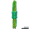
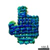
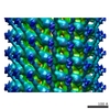
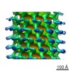
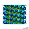
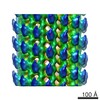
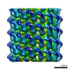
| Title | 126 helix bundle DNA nanostructure | |||||||||
 Map data Map data | hollow cylinder shaped 126 helix bundle DNA nanostructure | |||||||||
 Sample Sample |
| |||||||||
| Biological species | synthetic construct (others) | |||||||||
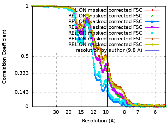
| Method | single particle reconstruction / cryo EM / Resolution: 9.8 Å | |||||||||
 Authors Authors | Kube M / Kohler F / Feigl E / Nagel-Yuksel B / Willner EM / Funke JJ / Gerling T / Stommer P / Honemann MN / Martin TG ...Kube M / Kohler F / Feigl E / Nagel-Yuksel B / Willner EM / Funke JJ / Gerling T / Stommer P / Honemann MN / Martin TG / Scheres SHW / Dietz H | |||||||||
| Funding support |  Germany, European Union, 2 items Germany, European Union, 2 items
| |||||||||
 Citation Citation | Journal: Nat Commun / Year: 2020 Title: Revealing the structures of megadalton-scale DNA complexes with nucleotide resolution. Authors: Massimo Kube / Fabian Kohler / Elija Feigl / Baki Nagel-Yüksel / Elena M Willner / Jonas J Funke / Thomas Gerling / Pierre Stömmer / Maximilian N Honemann / Thomas G Martin / Sjors H W ...Authors: Massimo Kube / Fabian Kohler / Elija Feigl / Baki Nagel-Yüksel / Elena M Willner / Jonas J Funke / Thomas Gerling / Pierre Stömmer / Maximilian N Honemann / Thomas G Martin / Sjors H W Scheres / Hendrik Dietz /  Abstract: The methods of DNA nanotechnology enable the rational design of custom shapes that self-assemble in solution from sets of DNA molecules. DNA origami, in which a long template DNA single strand is ...The methods of DNA nanotechnology enable the rational design of custom shapes that self-assemble in solution from sets of DNA molecules. DNA origami, in which a long template DNA single strand is folded by many short DNA oligonucleotides, can be employed to make objects comprising hundreds of unique DNA strands and thousands of base pairs, thus in principle providing many degrees of freedom for modelling complex objects of defined 3D shapes and sizes. Here, we address the problem of accurate structural validation of DNA objects in solution with cryo-EM based methodologies. By taking into account structural fluctuations, we can determine structures with improved detail compared to previous work. To interpret the experimental cryo-EM maps, we present molecular-dynamics-based methods for building pseudo-atomic models in a semi-automated fashion. Among other features, our data allows discerning details such as helical grooves, single-strand versus double-strand crossovers, backbone phosphate positions, and single-strand breaks. Obtaining this higher level of detail is a step forward that now allows designers to inspect and refine their designs with base-pair level interventions. | |||||||||
| History |
|
- Structure visualization
| Movie |
 Movie viewer Movie viewer |
|---|---|
| Structure viewer | EM map: SurfViewMolmilJmol/JSmol |
| Supplemental images |

- Downloads & links
-EMDB archive
| Map data | emd_11170.map.gz | 54.4 MB | EMDB map data format | |
|---|---|---|---|---|
| Header (meta data) | emd-11170-v30.xmlemd-11170.xml | 27.8 KB 27.8 KB | Display Display | EMDB header |
| FSC (resolution estimation) | emd_11170_fsc.xmlemd_11170_fsc_2.xmlemd_11170_fsc_3.xmlemd_11170_fsc_4.xmlemd_11170_fsc_5.xmlemd_11170_fsc_6.xmlemd_11170_fsc_7.xml | 10.7 KB 10.7 KB 10.8 KB 10.7 KB 10.7 KB 10.7 KB 10.7 KB | Display Display Display Display Display Display Display | FSC data file |
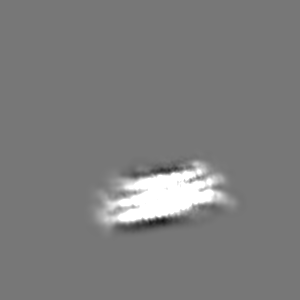
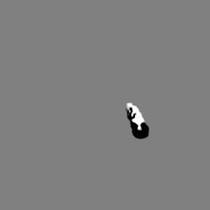
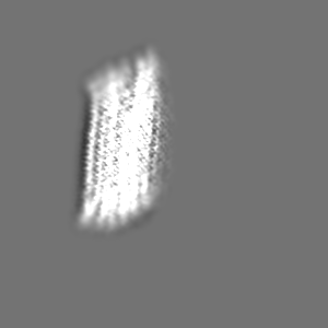
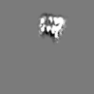
| Images | 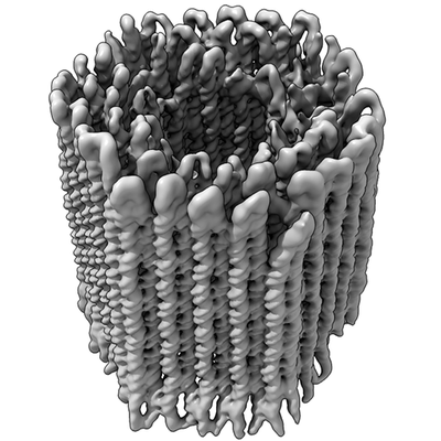 emd_11170.png emd_11170.png | 92.5 KB | ||
| Others | emd_11170_additional_1.map.gzemd_11170_additional_2.map.gzemd_11170_additional_3.map.gzemd_11170_additional_4.map.gzemd_11170_additional_5.map.gzemd_11170_additional_6.map.gzemd_11170_additional_7.map.gz | 5.9 MB 7.7 MB 5.9 MB 5.7 MB 5.7 MB 5.7 MB 6 MB | ||
| Archive directory |  http://ftp.pdbj.org/pub/emdb/structures/EMD-11170ftp://ftp.pdbj.org/pub/emdb/structures/EMD-11170 http://ftp.pdbj.org/pub/emdb/structures/EMD-11170ftp://ftp.pdbj.org/pub/emdb/structures/EMD-11170 | HTTPS FTP |
-Related structure data
| Related structure data |  7as5MC  7areC  7arqC  7artC  7arvC  7aryC C: citing same article ( M: atomic model generated by this map |
|---|---|
| Similar structure data |


-Links
| EMDB pages | EMDB (EBI/PDBe) / EMDataResource |
|---|---|
| Related items in Molecule of the Month |

-Map
| File | Download / File: emd_11170.map.gz / Format: CCP4 / Size: 103 MB / Type: IMAGE STORED AS FLOATING POINT NUMBER (4 BYTES) | ||||||||||||||||||||||||||||||||||||||||||||||||||||||||||||
|---|---|---|---|---|---|---|---|---|---|---|---|---|---|---|---|---|---|---|---|---|---|---|---|---|---|---|---|---|---|---|---|---|---|---|---|---|---|---|---|---|---|---|---|---|---|---|---|---|---|---|---|---|---|---|---|---|---|---|---|---|---|
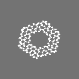
| Annotation | hollow cylinder shaped 126 helix bundle DNA nanostructure | ||||||||||||||||||||||||||||||||||||||||||||||||||||||||||||
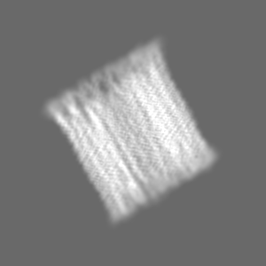
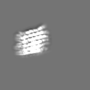
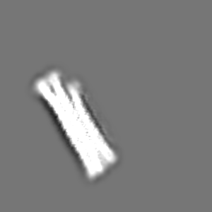
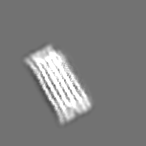
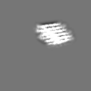
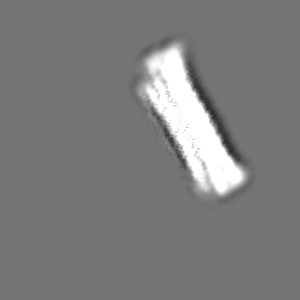
| Projections & slices | Image control
Images are generated by Spider. | ||||||||||||||||||||||||||||||||||||||||||||||||||||||||||||
| Voxel size | X=Y=Z: 2.78 Å | ||||||||||||||||||||||||||||||||||||||||||||||||||||||||||||
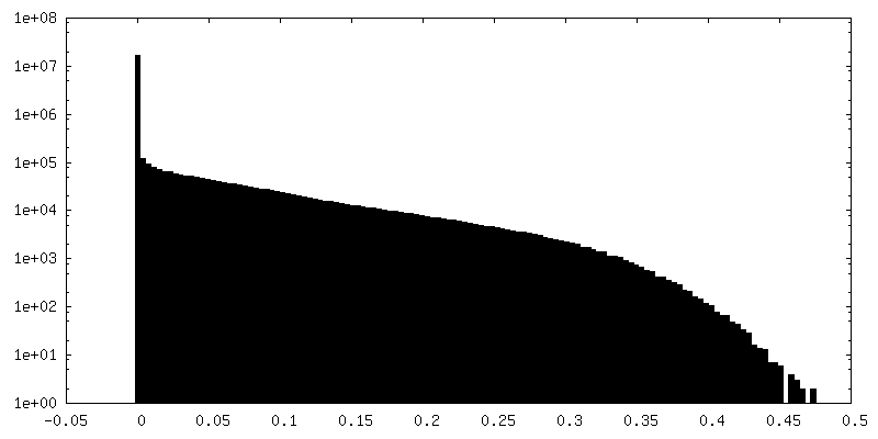
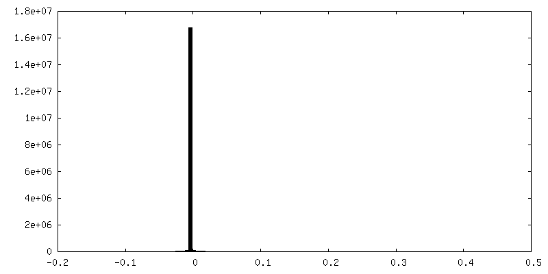
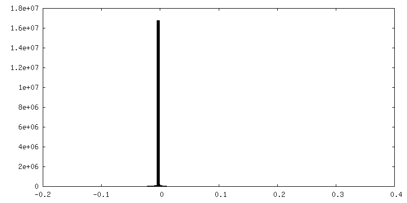
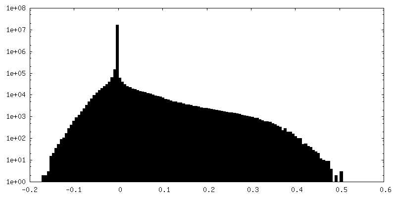
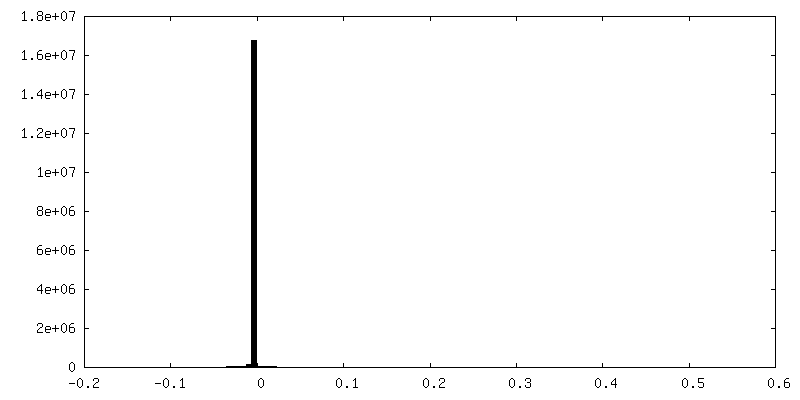
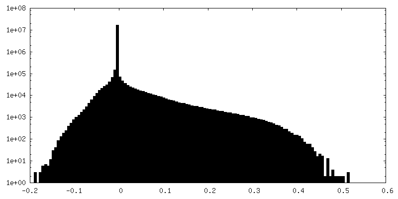
| Density |
| ||||||||||||||||||||||||||||||||||||||||||||||||||||||||||||
| Symmetry | Space group: 1 | ||||||||||||||||||||||||||||||||||||||||||||||||||||||||||||
| Details | EMDB XML:
CCP4 map header:
| ||||||||||||||||||||||||||||||||||||||||||||||||||||||||||||
 Z (Sec.)
 Y (Row.)
 X (Col.)


-Supplemental data
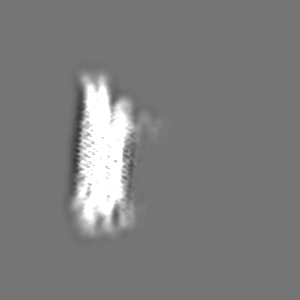
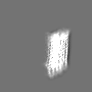
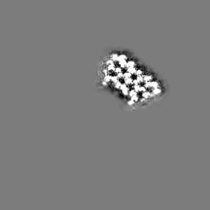
-Additional map: Body 4 of Multibody Refinement
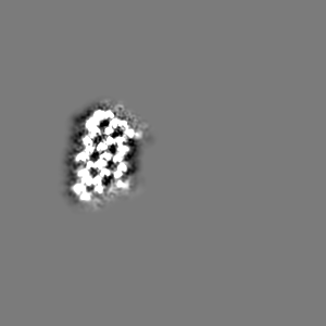
| File | emd_11170_additional_1.map | ||||||||||||
|---|---|---|---|---|---|---|---|---|---|---|---|---|---|
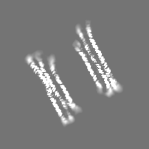
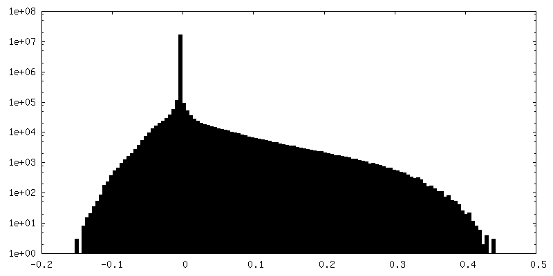
| Annotation | Body 4 of Multibody Refinement | ||||||||||||
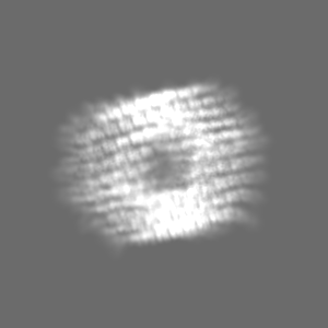
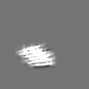
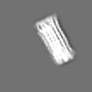
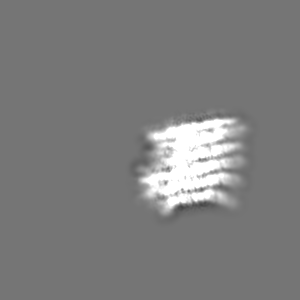
| Projections & Slices |
| ||||||||||||
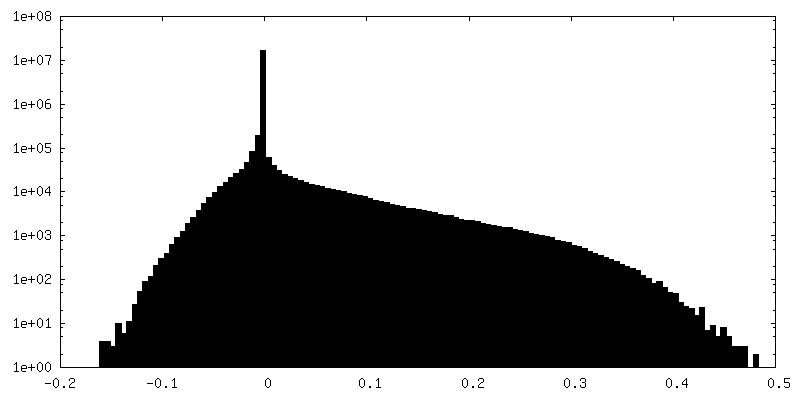
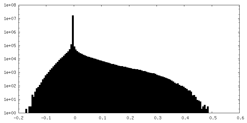
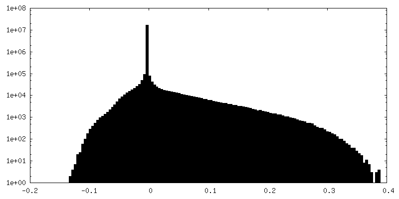
| Density Histograms |


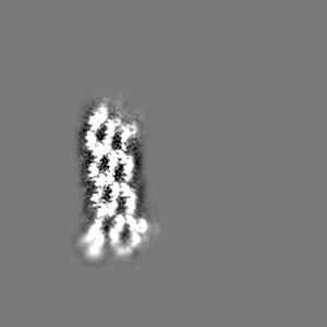
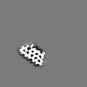
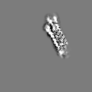
-Additional map: Composite map of Multibody Refinement (Relion) with 6 bodies
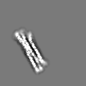
| File | emd_11170_additional_2.map | ||||||||||||
|---|---|---|---|---|---|---|---|---|---|---|---|---|---|
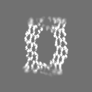
| Annotation | Composite map of Multibody Refinement (Relion) with 6 bodies | ||||||||||||
| Projections & Slices |
| ||||||||||||
| Density Histograms |
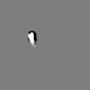
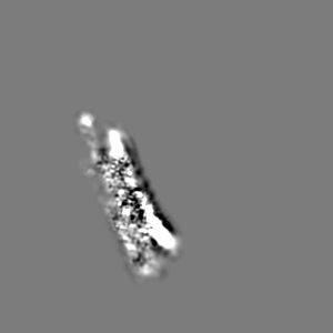
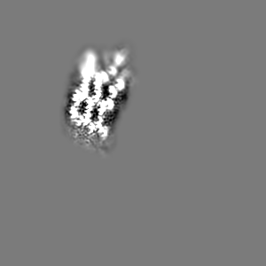
-Additional map: Body 3 of Multibody Refinement
| File | emd_11170_additional_3.map | ||||||||||||
|---|---|---|---|---|---|---|---|---|---|---|---|---|---|
| Annotation | Body 3 of Multibody Refinement | ||||||||||||
| Projections & Slices |
| ||||||||||||
| Density Histograms |

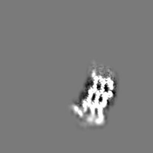
-Additional map: Body 1 of Multibody Refinement
| File | emd_11170_additional_4.map | ||||||||||||
|---|---|---|---|---|---|---|---|---|---|---|---|---|---|
| Annotation | Body 1 of Multibody Refinement | ||||||||||||
| Projections & Slices |
| ||||||||||||
| Density Histograms |


-Additional map: Body 2 of Multibody Refinement
| File | emd_11170_additional_5.map | ||||||||||||
|---|---|---|---|---|---|---|---|---|---|---|---|---|---|
| Annotation | Body 2 of Multibody Refinement | ||||||||||||
| Projections & Slices |
| ||||||||||||
| Density Histograms |
-Additional map: Body 5 of Multibody Refinement
| File | emd_11170_additional_6.map | ||||||||||||
|---|---|---|---|---|---|---|---|---|---|---|---|---|---|
| Annotation | Body 5 of Multibody Refinement | ||||||||||||
| Projections & Slices |
| ||||||||||||
| Density Histograms |

-Additional map: Body 6 of Multibody Refinement
| File | emd_11170_additional_7.map | ||||||||||||
|---|---|---|---|---|---|---|---|---|---|---|---|---|---|
| Annotation | Body 6 of Multibody Refinement | ||||||||||||
| Projections & Slices |
| ||||||||||||
| Density Histograms |


- Sample components
-Entire : 126 helix bundle
| Entire | Name: 126 helix bundle |
|---|---|
| Components |
|
-Supramolecule #1: 126 helix bundle
| Supramolecule | Name: 126 helix bundle / type: complex / ID: 1 / Parent: 0 Details: hollow cylinder-shaped multilayer DNA nanostructure with 126 helices arranged on a honey comb lattice |
|---|---|
| Source (natural) | Organism: synthetic construct (others) |
| Recombinant expression | Organism: synthetic construct (others) |
-Experimental details
-Structure determination
| Method | cryo EM |
|---|---|
 Processing Processing | single particle reconstruction |
| Aggregation state | particle |
-Sample preparation
| Buffer | pH: 8 |
|---|---|
| Grid | Model: C-flat-2/1 / Material: COPPER / Mesh: 400 / Support film - Material: CARBON / Support film - topology: HOLEY / Pretreatment - Type: GLOW DISCHARGE / Pretreatment - Atmosphere: AIR |
| Vitrification | Cryogen name: ETHANE / Chamber humidity: 100 % / Chamber temperature: 293 K / Instrument: FEI VITROBOT MARK IV |
| Details | monodisperse, 1200nM |
- Electron microscopy
| Microscope | FEI TITAN KRIOS |
|---|---|
| Specialist optics | Spherical aberration corrector: CEOS Cs corrector |
| Image recording | Film or detector model: FEI FALCON III (4k x 4k) / Detector mode: INTEGRATING / Digitization - Dimensions - Width: 4096 pixel / Digitization - Dimensions - Height: 4096 pixel / Number real images: 5005 / Average electron dose: 50.0 e/Å2 / Details: magnified pixel size 1.39 A |
| Electron beam | Acceleration voltage: 300 kV / Electron source:  FIELD EMISSION GUN FIELD EMISSION GUN |
| Electron optics | Calibrated defocus min: 0.3688 µm / Illumination mode: FLOOD BEAM / Imaging mode: BRIGHT FIELD / Cs: 0.01 mm / Nominal defocus max: 3.6056 µm / Nominal magnification: 47000 |
| Sample stage | Specimen holder model: FEI TITAN KRIOS AUTOGRID HOLDER / Cooling holder cryogen: NITROGEN |
| Experimental equipment |  Model: Titan Krios / Image courtesy: FEI Company |