 Movie
Movie Controller

[English] 日本語
 Yorodumi
- EMDB-1309: Human kinetochore-associated kinesin CENP-E visualized at 17 A re... -
+ Open data

- Basic information
| Entry | Database: EMDB / ID: EMD-1309 | |||||||||
|---|---|---|---|---|---|---|---|---|---|---|
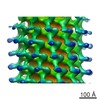
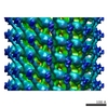
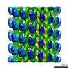
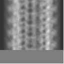
| Title | Human kinetochore-associated kinesin CENP-E visualized at 17 A resolution bound to microtubules. | |||||||||
 Map data Map data | Kinetochore-associated kinesin CENP-E motors bound to microtubules | |||||||||
 Sample Sample |
| |||||||||
| Biological species |   Homo sapiens (human) Homo sapiens (human) | |||||||||
| Method | helical reconstruction / cryo EM / Resolution: 17.0 Å | |||||||||
 Authors Authors | Neumann E / Garcia-Saez I / DeBonis S / Wade RH / Kozielski F / Conway JF | |||||||||
 Citation Citation | Journal: J Mol Biol / Year: 2006 Title: Human kinetochore-associated kinesin CENP-E visualized at 17 A resolution bound to microtubules. Authors: E Neumann / I Garcia-Saez / S DeBonis / R H Wade / F Kozielski / J F Conway /  Abstract: The highly dynamic process of cell division is effected, in part, by molecular motors that generate the forces necessary for its enactment. Several members of the kinesin superfamily of motor ...The highly dynamic process of cell division is effected, in part, by molecular motors that generate the forces necessary for its enactment. Several members of the kinesin superfamily of motor proteins are implicated in mitosis, such as CENP-E, which plays essential roles in cell division, including association with the kinetochore to stabilize attachment of chromosomes to microtubules prior to and during their separation. Neither the functional assembly state of CENP-E nor its direction of motion along the polar microtubule are certain. To determine the mode of interaction between CENP-E and microtubules, we have used cryo-electron microscopy to visualize CENP-E motor domains complexed with microtubules and calculated a density map of the complex to 17 A resolution by combining helical and single-particle reconstruction methods. The interface between the motor domain and microtubules was modeled by docking atomic-resolution models of the subunits into the cryoEM density map. Our results support a plus end motion for CENP-E, consistent with features of the crystallographic structure. Despite considerable functional differences from the monomeric transporter kinesin KIF1A and the oppositely directed ncd kinesin, CENP-E appears to share many features of the intermolecular interactions, suggesting that differences in motor function are governed by small variations in the loops at the microtubule interface. | |||||||||
| History |
|
- Structure visualization
| Movie |
 Movie viewer Movie viewer |
|---|---|
| Structure viewer | EM map: SurfViewMolmilJmol/JSmol |
| Supplemental images |
 UCSF Chimera
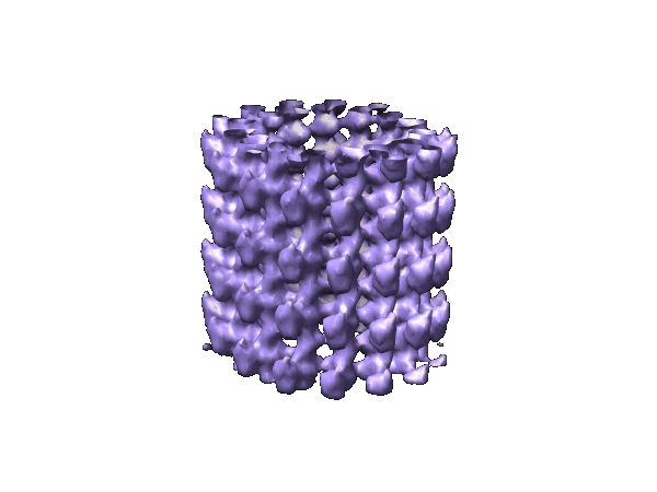
- Downloads & links
-EMDB archive
| Map data | emd_1309.map.gz | 1 MB | EMDB map data format | |
|---|---|---|---|---|
| Header (meta data) | emd-1309-v30.xmlemd-1309.xml | 10.1 KB 10.1 KB | Display Display | EMDB header |
| Images |  1309.gif 1309.gif | 54.6 KB | ||
| Archive directory |  http://ftp.pdbj.org/pub/emdb/structures/EMD-1309ftp://ftp.pdbj.org/pub/emdb/structures/EMD-1309 http://ftp.pdbj.org/pub/emdb/structures/EMD-1309ftp://ftp.pdbj.org/pub/emdb/structures/EMD-1309 | HTTPS FTP |
-Related structure data
| Similar structure data |
|---|
-Links
| EMDB pages | EMDB (EBI/PDBe) / EMDataResource |
|---|
-Map
| File | Download / File: emd_1309.map.gz / Format: CCP4 / Size: 2 MB / Type: IMAGE STORED AS SIGNED BYTE | ||||||||||||||||||||||||||||||||||||||||||||||||||||||||||||||||||||
|---|---|---|---|---|---|---|---|---|---|---|---|---|---|---|---|---|---|---|---|---|---|---|---|---|---|---|---|---|---|---|---|---|---|---|---|---|---|---|---|---|---|---|---|---|---|---|---|---|---|---|---|---|---|---|---|---|---|---|---|---|---|---|---|---|---|---|---|---|---|
| Annotation | Kinetochore-associated kinesin CENP-E motors bound to microtubules | ||||||||||||||||||||||||||||||||||||||||||||||||||||||||||||||||||||
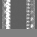
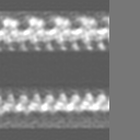
| Projections & slices | Image control
Images are generated by Spider. | ||||||||||||||||||||||||||||||||||||||||||||||||||||||||||||||||||||
| Voxel size | X=Y=Z: 3.56 Å | ||||||||||||||||||||||||||||||||||||||||||||||||||||||||||||||||||||
| Density |
| ||||||||||||||||||||||||||||||||||||||||||||||||||||||||||||||||||||
| Symmetry | Space group: 1 | ||||||||||||||||||||||||||||||||||||||||||||||||||||||||||||||||||||
| Details | EMDB XML:
CCP4 map header:
| ||||||||||||||||||||||||||||||||||||||||||||||||||||||||||||||||||||
 Y (Sec.)
 X (Row.)
 Z (Col.)


-Supplemental data
- Sample components
-Entire : Human Kinetochore-associated Kinesin CENP-E bound to Microtubules
| Entire | Name: Human Kinetochore-associated Kinesin CENP-E bound to Microtubules |
|---|---|
| Components |
|
-Supramolecule #1000: Human Kinetochore-associated Kinesin CENP-E bound to Microtubules
| Supramolecule | Name: Human Kinetochore-associated Kinesin CENP-E bound to Microtubules type: sample / ID: 1000 Oligomeric state: One CENP-E motor domain binds to a tubulin heterodimer Number unique components: 2 |
|---|
-Macromolecule #1: Polymerized Tubulin
| Macromolecule | Name: Polymerized Tubulin / type: protein_or_peptide / ID: 1 / Name.synonym: Microtubules / Details: cat. no. TL238, Cytoskeleton Inc, Denver CO, USA / Number of copies: 2 / Oligomeric state: Dimer / Recombinant expression: No / Database: NCBI |
|---|---|
| Source (natural) | Organism: |
-Macromolecule #2: Human kinetochore-associated kinesin CENP-E motors
| Macromolecule | Name: Human kinetochore-associated kinesin CENP-E motors / type: protein_or_peptide / ID: 2 / Name.synonym: Mitotic Kinesins / Details: Monomeric Form / Number of copies: 1 / Oligomeric state: Monomer / Recombinant expression: Yes |
|---|---|
| Source (natural) | Organism: Homo sapiens (human) / synonym: Human |
| Recombinant expression | Organism:  |
-Experimental details
-Structure determination
| Method | cryo EM |
|---|---|
 Processing Processing | helical reconstruction |
| Aggregation state | filament |
-Sample preparation
| Buffer | pH: 6 Details: 20 mM sodium phosphate, 50 mM NaCl, 1 mM MgCl2, 1 mM EGTA |
|---|---|
| Grid | Details: Holey carbon grid |
| Vitrification | Cryogen name: ETHANE / Details: Vitrification carried out in nitrogen atmosphere / Method: Blot for 2 seconds before plunging |
- Electron microscopy
| Microscope | FEI/PHILIPS CM200T |
|---|---|
| Image recording | Category: FILM / Film or detector model: KODAK SO-163 FILM / Digitization - Scanner: OTHER / Digitization - Sampling interval: 7 µm / Number real images: 21 / Bits/pixel: 8 |
| Electron beam | Acceleration voltage: 200 kV / Electron source: LAB6 |
| Electron optics | Calibrated magnification: 39325 / Illumination mode: OTHER / Imaging mode: BRIGHT FIELD / Cs: 2 mm / Nominal defocus max: 4.0 µm / Nominal defocus min: 1.2 µm / Nominal magnification: 38000 |
| Sample stage | Specimen holder: Eucentric / Specimen holder model: GATAN LIQUID NITROGEN |
-Image processing
| Final reconstruction | Algorithm: OTHER / Resolution.type: BY AUTHOR / Resolution: 17.0 Å / Resolution method: FSC 0.33 CUT-OFF / Software - Name: MRC and Spider |
|---|---|
| CTF correction | Details: CTFMIX |
-Atomic model buiding 1
| Software | Name: Situs, URO |
|---|---|
| Details | The cryoEM map and the atomic structures of CENP-E and the tubulin dimer were visualized together using the program "O". An initial manual fit was straightforward, and subsequently refined using CoLoRes from the SITUS package, and URO. |
| Refinement | Protocol: RIGID BODY FIT |
