 Movie
Movie Controller

[English] 日本語
 Yorodumi
- EMDB-1029: Image reconstructions of microtubules decorated with monomeric an... -
+ Open data

- Basic information
| Entry | Database: EMDB / ID: EMD-1029 | |||||||||
|---|---|---|---|---|---|---|---|---|---|---|
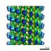
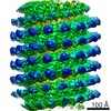
| Title | Image reconstructions of microtubules decorated with monomeric and dimeric kinesins: comparison with x-ray structure and implications for motility. | |||||||||
 Map data Map data | rat kinesin monomer AMP-PNP state | |||||||||
 Sample Sample |
| |||||||||
| Biological species |  | |||||||||
| Method | helical reconstruction / cryo EM / negative staining / Resolution: 25.0 Å | |||||||||
 Authors Authors | Hoenger A | |||||||||
 Citation Citation | Journal: J Cell Biol / Year: 1998 Title: Image reconstructions of microtubules decorated with monomeric and dimeric kinesins: comparison with x-ray structure and implications for motility. Authors: A Hoenger / S Sack / M Thormählen / A Marx / J Müller / H Gross / E Mandelkow /  Abstract: We have decorated microtubules with monomeric and dimeric kinesin constructs, studied their structure by cryoelectron microscopy and three-dimensional image reconstruction, and compared the results ...We have decorated microtubules with monomeric and dimeric kinesin constructs, studied their structure by cryoelectron microscopy and three-dimensional image reconstruction, and compared the results with the x-ray crystal structure of monomeric and dimeric kinesin. A monomeric kinesin construct (rK354, containing only a short neck helix insufficient for coiled-coil formation) decorates microtubules with a stoichiometry of one kinesin head per tubulin subunit (alpha-beta-heterodimer). The orientation of the kinesin head (an anterograde motor) on the microtubule surface is similar to that of ncd (a retrograde motor). A longer kinesin construct (rK379) forms a dimer because of the longer neck helix forming a coiled-coil. Unexpectedly, this construct also decorates the microtubule with a stoichiometry of one head per tubulin subunit, and the orientation is similar to that of the monomeric construct. This means that the interaction with microtubules causes the two heads of a kinesin dimer to separate sufficiently so that they can bind to two different tubulin subunits. This result is in contrast to recent models and can be explained by assuming that the tubulin-kinesin interaction is antagonistic to the coiled-coil interaction within a kinesin dimer. | |||||||||
| History |
|
- Structure visualization
| Movie |
 Movie viewer Movie viewer |
|---|---|
| Structure viewer | EM map: SurfViewMolmilJmol/JSmol |
| Supplemental images |
 UCSF Chimera
- Downloads & links
-EMDB archive
| Map data | emd_1029.map.gz | 2.7 MB | EMDB map data format | |
|---|---|---|---|---|
| Header (meta data) | emd-1029-v30.xmlemd-1029.xml | 10 KB 10 KB | Display Display | EMDB header |
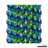
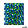
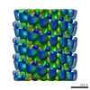
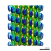
| Images | 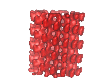 1029.gif 1029.gif | 29 KB | ||
| Archive directory |  http://ftp.pdbj.org/pub/emdb/structures/EMD-1029ftp://ftp.pdbj.org/pub/emdb/structures/EMD-1029 http://ftp.pdbj.org/pub/emdb/structures/EMD-1029ftp://ftp.pdbj.org/pub/emdb/structures/EMD-1029 | HTTPS FTP |
-Related structure data
-Links
| EMDB pages | EMDB (EBI/PDBe) / EMDataResource |
|---|
-Map
| File | Download / File: emd_1029.map.gz / Format: CCP4 / Size: 3.7 MB / Type: IMAGE STORED AS FLOATING POINT NUMBER (4 BYTES) | ||||||||||||||||||||||||||||||||||||||||||||||||||||||||||||
|---|---|---|---|---|---|---|---|---|---|---|---|---|---|---|---|---|---|---|---|---|---|---|---|---|---|---|---|---|---|---|---|---|---|---|---|---|---|---|---|---|---|---|---|---|---|---|---|---|---|---|---|---|---|---|---|---|---|---|---|---|---|
| Annotation | rat kinesin monomer AMP-PNP state | ||||||||||||||||||||||||||||||||||||||||||||||||||||||||||||
| Projections & slices | Image control
Images are generated by Spider. | ||||||||||||||||||||||||||||||||||||||||||||||||||||||||||||
| Voxel size | X=Y=Z: 5.714 Å | ||||||||||||||||||||||||||||||||||||||||||||||||||||||||||||
| Density |
| ||||||||||||||||||||||||||||||||||||||||||||||||||||||||||||
| Symmetry | Space group: 1 | ||||||||||||||||||||||||||||||||||||||||||||||||||||||||||||
| Details | EMDB XML:
CCP4 map header:
| ||||||||||||||||||||||||||||||||||||||||||||||||||||||||||||
 Z (Sec.)
 Y (Row.)
 X (Col.)


-Supplemental data
- Sample components
-Entire : rat kinesin motor domains complexed to microtubules
| Entire | Name: rat kinesin motor domains complexed to microtubules |
|---|---|
| Components |
|
-Supramolecule #1000: rat kinesin motor domains complexed to microtubules
| Supramolecule | Name: rat kinesin motor domains complexed to microtubules / type: sample / ID: 1000 / Oligomeric state: monomer / Number unique components: 2 |
|---|
-Macromolecule #1: rat kinesin
| Macromolecule | Name: rat kinesin / type: protein_or_peptide / ID: 1 / Name.synonym: molecular motor / Number of copies: 1 / Recombinant expression: Yes |
|---|---|
| Source (natural) | Organism: |
| Recombinant expression | Organism:  |
-Macromolecule #2: tubulin
| Macromolecule | Name: tubulin / type: protein_or_peptide / ID: 2 / Name.synonym: microtubules / Number of copies: 1 / Recombinant expression: Yes |
|---|---|
| Source (natural) | Organism: |
| Recombinant expression | Organism: |
-Experimental details
-Structure determination
| Method | negative staining, cryo EM |
|---|---|
 Processing Processing | helical reconstruction |
| Aggregation state | filament |
-Sample preparation
| Concentration | 0.5 mg/mL |
|---|---|
| Buffer | pH: 6.8 / Details: Pipes 80mM, MgCl 1mM, GTP 1mM, Taxol 20uM, DMSO 5% |
| Staining | Type: NEGATIVE / Details: ice-embedded |
| Grid | Details: holey grids |
| Vitrification | Cryogen name: ETHANE / Chamber temperature: 93 K / Instrument: HOMEMADE PLUNGER / Details: Vitrification instrument: self made |
- Electron microscopy
| Microscope | FEI/PHILIPS CM12 |
|---|---|
| Temperature | Average: 95 K |
| Image recording | Category: FILM / Film or detector model: AGFA SCIENTA FILM / Digitization - Scanner: EMIL 10 / Digitization - Sampling interval: 20 µm / Number real images: 10 / Average electron dose: 5 e/Å2 / Bits/pixel: 16 |
| Tilt angle min | 0 |
| Tilt angle max | 0 |
| Electron beam | Acceleration voltage: 100 kV / Electron source: LAB6 |
| Electron optics | Illumination mode: FLOOD BEAM / Imaging mode: BRIGHT FIELD / Nominal defocus max: 1.6 µm / Nominal defocus min: 1.0 µm / Nominal magnification: 35000 |
| Sample stage | Specimen holder: side-entry / Specimen holder model: GATAN LIQUID NITROGEN |
-Image processing
| Final reconstruction | Applied symmetry - Helical parameters - Axial symmetry: C1 (asymmetric) Algorithm: OTHER / Resolution.type: BY AUTHOR / Resolution: 25.0 Å / Resolution method: FSC 0.5 CUT-OFF / Software - Name: Phoelix, Suprim Details: Final maps from 20 averaged datasets = 10 helical tubes |
|---|
