 Movie
Movie Controller

[English] 日本語
 Yorodumi
- EMDB-1035: A new look at the microtubule binding patterns of dimeric kinesins. -
+ Open data

- Basic information
| Entry | Database: EMDB / ID: EMD-1035 | |||||||||
|---|---|---|---|---|---|---|---|---|---|---|
| Title | A new look at the microtubule binding patterns of dimeric kinesins. | |||||||||
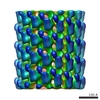
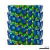
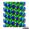
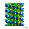
 Map data Map data | Neurospora crassa kinesin monomer (nK343) AMP-PNP state | |||||||||
 Sample Sample |
| |||||||||
| Biological species |  Neurospora crassa (fungus) Neurospora crassa (fungus) | |||||||||
| Method | helical reconstruction / cryo EM / negative staining / Resolution: 25.0 Å | |||||||||
 Authors Authors | Hoenger A | |||||||||
 Citation Citation | Journal: J Mol Biol / Year: 2000 Title: A new look at the microtubule binding patterns of dimeric kinesins. Authors: A Hoenger / M Thormählen / R Diaz-Avalos / M Doerhoefer / K N Goldie / J Müller / E Mandelkow /  Abstract: The interactions of monomeric and dimeric kinesin and ncd constructs with microtubules have been investigated using cryo-electron microscopy (cryo-EM) and several biochemical methods. There is a good ...The interactions of monomeric and dimeric kinesin and ncd constructs with microtubules have been investigated using cryo-electron microscopy (cryo-EM) and several biochemical methods. There is a good consensus on the structure of dimeric ncd when bound to a tubulin dimer showing one head attached directly to tubulin, and the second head tethered to the first. However, the 3D maps of dimeric kinesin motor domains are still quite controversial and leave room for different interpretations. Here we reinvestigated the microtubule binding patterns of dimeric kinesins by cryo-EM and digital 3D reconstruction under different nucleotide conditions and different motor:tubulin ratios, and determined the molecular mass of motor-tubulin complexes by STEM. Both methods revealed complementary results. We found that the ratio of bound kinesin motor-heads to alphabeta-tubulin dimers was never reaching above 1.5 irrespective of the initial mixing ratios. It appears that each kinesin dimer occupies two microtubule-binding sites, provided that there is a free one nearby. Thus the appearances of different image reconstructions can be explained by non-specific excess binding of motor heads. Consequently, the use of different apparent density distributions for docking the X-ray structures onto the microtubule surface leads to different and mutually exclusive models. We propose that in conditions of stoichiometric binding the two heads of a kinesin dimer separate and bind to different tubulin subunits. This is in contrast to ncd where the two heads remain tightly attached on the microtubule surface. Using dimeric kinesin molecules crosslinked in their neck domain we also found that they stabilize protofilaments axially, but not laterally, which is a strong indication that the two heads of the dimers bind along one protofilament, rather than laterally bridging two protofilaments. A molecular walking model based on these results summarizes our conclusions and illustrates the implications of symmetry for such models. | |||||||||
| History |
|
- Structure visualization
| Movie |
 Movie viewer Movie viewer |
|---|---|
| Structure viewer | EM map: SurfViewMolmilJmol/JSmol |
| Supplemental images |
 UCSF Chimera
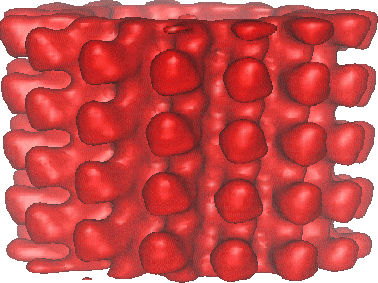
- Downloads & links
-EMDB archive
| Map data | emd_1035.map.gz | 2.7 MB | EMDB map data format | |
|---|---|---|---|---|
| Header (meta data) | emd-1035-v30.xmlemd-1035.xml | 9.6 KB 9.6 KB | Display Display | EMDB header |
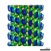
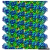
| Images |  1035.gif 1035.gif | 61.7 KB | ||
| Archive directory |  http://ftp.pdbj.org/pub/emdb/structures/EMD-1035ftp://ftp.pdbj.org/pub/emdb/structures/EMD-1035 http://ftp.pdbj.org/pub/emdb/structures/EMD-1035ftp://ftp.pdbj.org/pub/emdb/structures/EMD-1035 | HTTPS FTP |
-Related structure data
-Links
| EMDB pages | EMDB (EBI/PDBe) / EMDataResource |
|---|
-Map
| File | Download / File: emd_1035.map.gz / Format: CCP4 / Size: 3.7 MB / Type: IMAGE STORED AS FLOATING POINT NUMBER (4 BYTES) | ||||||||||||||||||||||||||||||||||||||||||||||||||||||||||||
|---|---|---|---|---|---|---|---|---|---|---|---|---|---|---|---|---|---|---|---|---|---|---|---|---|---|---|---|---|---|---|---|---|---|---|---|---|---|---|---|---|---|---|---|---|---|---|---|---|---|---|---|---|---|---|---|---|---|---|---|---|---|
| Annotation | Neurospora crassa kinesin monomer (nK343) AMP-PNP state | ||||||||||||||||||||||||||||||||||||||||||||||||||||||||||||
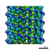
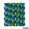
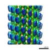
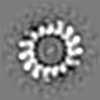
| Projections & slices | Image control
Images are generated by Spider. | ||||||||||||||||||||||||||||||||||||||||||||||||||||||||||||
| Voxel size | X=Y=Z: 5.676 Å | ||||||||||||||||||||||||||||||||||||||||||||||||||||||||||||
| Density |
| ||||||||||||||||||||||||||||||||||||||||||||||||||||||||||||
| Symmetry | Space group: 1 | ||||||||||||||||||||||||||||||||||||||||||||||||||||||||||||
| Details | EMDB XML:
CCP4 map header:
| ||||||||||||||||||||||||||||||||||||||||||||||||||||||||||||
 Z (Sec.)
 Y (Row.)
 X (Col.)


-Supplemental data
- Sample components
-Entire : neurospora crassa kinesin monomer
| Entire | Name: neurospora crassa kinesin monomer |
|---|---|
| Components |
|
-Supramolecule #1000: neurospora crassa kinesin monomer
| Supramolecule | Name: neurospora crassa kinesin monomer / type: sample / ID: 1000 / Oligomeric state: monomer / Number unique components: 2 |
|---|
-Macromolecule #1: neurospora kinesin
| Macromolecule | Name: neurospora kinesin / type: protein_or_peptide / ID: 1 / Name.synonym: molecular motor / Number of copies: 1 / Oligomeric state: monomer / Recombinant expression: Yes / Database: NCBI |
|---|---|
| Source (natural) | Organism: Neurospora crassa (fungus) / synonym: neurospora kinesin |
-Macromolecule #2: tubulin
| Macromolecule | Name: tubulin / type: protein_or_peptide / ID: 2 / Name.synonym: microtubules / Number of copies: 1 / Oligomeric state: hetero dimer / Recombinant expression: No / Database: NCBI |
|---|---|
| Source (natural) | Organism: Neurospora crassa (fungus) / synonym: neurospora kinesin / Tissue: brain / Cell: neuronal cells / Location in cell: cytoplasm |
-Experimental details
-Structure determination
| Method | negative staining, cryo EM |
|---|---|
 Processing Processing | helical reconstruction |
| Aggregation state | filament |
-Sample preparation
| Concentration | 0.5 mg/mL |
|---|---|
| Buffer | pH: 6.9 Details: Pipes 20mM, 50 mM NaCl, 5mM mgcl,1mM Mg-ATP, 20um taxol. |
| Staining | Type: NEGATIVE / Details: ice-embedded |
| Grid | Details: holey grids |
| Vitrification | Cryogen name: ETHANE / Chamber temperature: 93 K / Instrument: HOMEMADE PLUNGER / Details: Vitrification instrument: self made |
- Electron microscopy
| Microscope | FEI/PHILIPS CM120T |
|---|---|
| Image recording | Category: FILM / Film or detector model: KODAK SO-163 FILM / Digitization - Scanner: ZEISS SCAI / Digitization - Sampling interval: 21 µm / Number real images: 10 / Average electron dose: 5 e/Å2 / Bits/pixel: 16 |
| Tilt angle min | 0 |
| Tilt angle max | 0 |
| Electron beam | Acceleration voltage: 100 kV / Electron source: LAB6 |
| Electron optics | Illumination mode: FLOOD BEAM / Imaging mode: BRIGHT FIELD / Nominal defocus max: 2.0 µm / Nominal defocus min: 1.5 µm / Nominal magnification: 37000 |
| Sample stage | Specimen holder: side entry / Specimen holder model: GATAN LIQUID NITROGEN |
-Image processing
| Final reconstruction | Applied symmetry - Helical parameters - Axial symmetry: C1 (asymmetric) Algorithm: OTHER / Resolution.type: BY AUTHOR / Resolution: 25.0 Å / Resolution method: FSC 0.5 CUT-OFF / Software - Name: Phoelix, Suprim Details: Final maps from 20 averaged datasets = 10 helical tubes |
|---|
