 Movie
Movie Controller

[English] 日本語
 Yorodumi
- EMDB-10639: Structure of ryanodine receptor 1 in apo state in native membrane... -
+ Open data

- Basic information
| Entry | Database: EMDB / ID: EMD-10639 | |||||||||
|---|---|---|---|---|---|---|---|---|---|---|
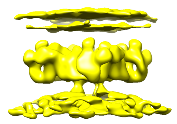
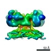
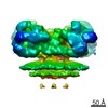
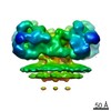
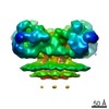
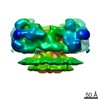
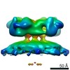
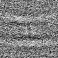
| Title | Structure of ryanodine receptor 1 in apo state in native membrane with the T-tubule membrane | |||||||||
 Map data Map data | Structure of ryanodine receptor 1 in app state in native membrane with the T-tubule membrane | |||||||||
 Sample Sample |
| |||||||||
| Biological species |  | |||||||||
| Method | subtomogram averaging / cryo EM / Resolution: 38.0 Å | |||||||||
 Authors Authors | Chen W / Kudryashev M | |||||||||
| Funding support |  Germany, 1 items Germany, 1 items
| |||||||||
 Citation Citation | Journal: EMBO Rep / Year: 2020 Title: Structure of RyR1 in native membranes. Authors: Wenbo Chen / Mikhail Kudryashev / Abstract: Ryanodine receptor 1 (RyR1) mediates excitation-contraction coupling by releasing Ca from sarcoplasmic reticulum (SR) to the cytoplasm of skeletal muscle cells. RyR1 activation is regulated by ...Ryanodine receptor 1 (RyR1) mediates excitation-contraction coupling by releasing Ca from sarcoplasmic reticulum (SR) to the cytoplasm of skeletal muscle cells. RyR1 activation is regulated by several proteins from both the cytoplasm and lumen of the SR. Here, we report the structure of RyR1 from native SR membranes in closed and open states. Compared to the previously reported structures of purified RyR1, our structure reveals helix-like densities traversing the bilayer approximately 5 nm from the RyR1 transmembrane domain and sarcoplasmic extensions linking RyR1 to a putative calsequestrin network. We document the primary conformation of RyR1 in situ and its structural variations. The activation of RyR1 is associated with changes in membrane curvature and movement in the sarcoplasmic extensions. Our results provide structural insight into the mechanism of RyR1 in its native environment. | |||||||||
| History |
|
- Structure visualization
| Movie |
 Movie viewer Movie viewer |
|---|---|
| Structure viewer | EM map: SurfViewMolmilJmol/JSmol |
| Supplemental images |
- Downloads & links
-EMDB archive
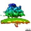
| Map data | emd_10639.map.gz | 26.2 MB | EMDB map data format | |
|---|---|---|---|---|
| Header (meta data) | emd-10639-v30.xmlemd-10639.xml | 13.7 KB 13.7 KB | Display Display | EMDB header |
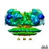
| Images |  emd_10639.png emd_10639.png | 116.5 KB | ||
| Masks | emd_10639_msk_1.map | 30.5 MB | Mask map | |
| Others | emd_10639_half_map_1.map.gzemd_10639_half_map_2.map.gz | 28.4 MB 28.4 MB | ||
| Archive directory |  http://ftp.pdbj.org/pub/emdb/structures/EMD-10639ftp://ftp.pdbj.org/pub/emdb/structures/EMD-10639 http://ftp.pdbj.org/pub/emdb/structures/EMD-10639ftp://ftp.pdbj.org/pub/emdb/structures/EMD-10639 | HTTPS FTP |
-Related structure data
| Related structure data | C: citing same article ( |
|---|---|
| Similar structure data | |
| EM raw data | EMPIAR-10349 (Title: Cryo Electron Tomograms of Membrane Fractions of Rabbit Skeletal Muscle for Structural Determination of RyR1 in SR Vesicles Data size: 254.2 Data #1: 82 tilt series (frames aligned my motioncorr2) in .st stacks (mrc format for imod) including .tlt, .xf and .defocus files [tilt series]) |

-Links
| EMDB pages | EMDB (EBI/PDBe) / EMDataResource |
|---|
-Map
| File | Download / File: emd_10639.map.gz / Format: CCP4 / Size: 30.5 MB / Type: IMAGE STORED AS FLOATING POINT NUMBER (4 BYTES) | ||||||||||||||||||||||||||||||||||||||||||||||||||||||||||||
|---|---|---|---|---|---|---|---|---|---|---|---|---|---|---|---|---|---|---|---|---|---|---|---|---|---|---|---|---|---|---|---|---|---|---|---|---|---|---|---|---|---|---|---|---|---|---|---|---|---|---|---|---|---|---|---|---|---|---|---|---|---|
| Annotation | Structure of ryanodine receptor 1 in app state in native membrane with the T-tubule membrane | ||||||||||||||||||||||||||||||||||||||||||||||||||||||||||||
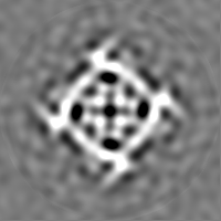
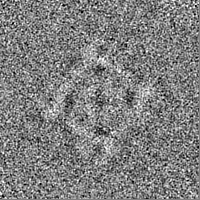
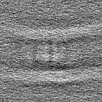
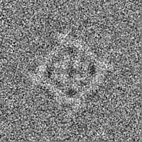
| Projections & slices | Image control
Images are generated by Spider. | ||||||||||||||||||||||||||||||||||||||||||||||||||||||||||||
| Voxel size | X=Y=Z: 2.7 Å | ||||||||||||||||||||||||||||||||||||||||||||||||||||||||||||
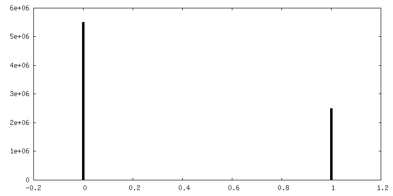
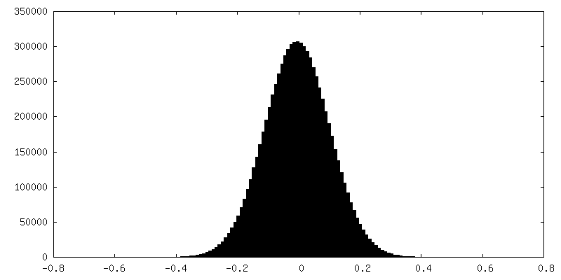
| Density |
| ||||||||||||||||||||||||||||||||||||||||||||||||||||||||||||
| Symmetry | Space group: 1 | ||||||||||||||||||||||||||||||||||||||||||||||||||||||||||||
| Details | EMDB XML:
CCP4 map header:
| ||||||||||||||||||||||||||||||||||||||||||||||||||||||||||||
 Z (Sec.)
 Y (Row.)
 X (Col.)


-Supplemental data
-Mask #1
| File | emd_10639_msk_1.map | ||||||||||||
|---|---|---|---|---|---|---|---|---|---|---|---|---|---|
| Projections & Slices |
| ||||||||||||
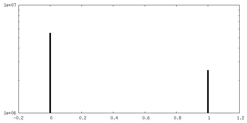
| Density Histograms |


-Half map: Structure of ryanodine receptor 1 in app state...
| File | emd_10639_half_map_1.map | ||||||||||||
|---|---|---|---|---|---|---|---|---|---|---|---|---|---|
| Annotation | Structure of ryanodine receptor 1 in app state in native membrane with the T-tubule membrane (half map 1) | ||||||||||||
| Projections & Slices |
| ||||||||||||
| Density Histograms |

-Half map: Structure of ryanodine receptor 1 in app state...
| File | emd_10639_half_map_2.map | ||||||||||||
|---|---|---|---|---|---|---|---|---|---|---|---|---|---|
| Annotation | Structure of ryanodine receptor 1 in app state in native membrane with the T-tubule membrane (half map 2) | ||||||||||||
| Projections & Slices |
| ||||||||||||
| Density Histograms |


- Sample components
-Entire : Rabbit ryanodine receptor 1 in apo state in native membrane
| Entire | Name: Rabbit ryanodine receptor 1 in apo state in native membrane |
|---|---|
| Components |
|
-Supramolecule #1: Rabbit ryanodine receptor 1 in apo state in native membrane
| Supramolecule | Name: Rabbit ryanodine receptor 1 in apo state in native membrane type: complex / ID: 1 / Parent: 0 |
|---|---|
| Source (natural) | Organism: |
-Experimental details
-Structure determination
| Method | cryo EM |
|---|---|
 Processing Processing | subtomogram averaging |
| Aggregation state | tissue |
-Sample preparation
| Buffer | pH: 7.1 |
|---|---|
| Vitrification | Cryogen name: ETHANE |
- Electron microscopy
| Microscope | FEI TITAN KRIOS |
|---|---|
| Image recording | Film or detector model: GATAN K2 SUMMIT (4k x 4k) / Detector mode: COUNTING / Average electron dose: 2.0 e/Å2 |
| Electron beam | Acceleration voltage: 300 kV / Electron source:  FIELD EMISSION GUN FIELD EMISSION GUN |
| Electron optics | Illumination mode: FLOOD BEAM / Imaging mode: BRIGHT FIELD |
| Experimental equipment |  Model: Titan Krios / Image courtesy: FEI Company |
