 Movie
Movie Controller

[English] 日本語
 Yorodumi
- PDB-1qu7: FOUR HELICAL-BUNDLE STRUCTURE OF THE CYTOPLASMIC DOMAIN OF A SERI... -
+ Open data

- Basic information
| Entry | Database: PDB / ID: 1qu7 | ||||||
|---|---|---|---|---|---|---|---|
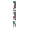
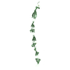
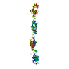
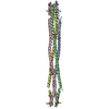
| Title | FOUR HELICAL-BUNDLE STRUCTURE OF THE CYTOPLASMIC DOMAIN OF A SERINE CHEMOTAXIS RECEPTOR | ||||||
 Components Components | METHYL-ACCEPTING CHEMOTAXIS PROTEIN I | ||||||
 Keywords Keywords | SIGNALING PROTEIN / SERINE / CHEMOTAXIS / FOUR HELICAL-BUNDLE | ||||||
| Function / homology |  Function and homology information Function and homology informationdetection of chemical stimulus / methyl accepting chemotaxis protein complex / regulation of bacterial-type flagellum-dependent cell motility / cell motility / cellular response to amino acid stimulus / chemotaxis / transmembrane signaling receptor activity / signal transduction / identical protein binding / plasma membrane Similarity search - Function | ||||||
| Biological species |  | ||||||
| Method |  X-RAY DIFFRACTION / SYNCHROTRON / Resolution: 2.6 Å X-RAY DIFFRACTION / SYNCHROTRON / Resolution: 2.6 Å | ||||||
 Authors Authors | Kim, K.K. / Yokota, H. / Kim, S.-H. | ||||||
 Citation Citation | Journal: Nature / Year: 1999 Title: Four-helical-bundle structure of the cytoplasmic domain of a serine chemotaxis receptor. Authors: Kim, K.K. / Yokota, H. / Kim, S.H. | ||||||
| History |
|
- Structure visualization
| Structure viewer | Molecule: MolmilJmol/JSmol |
|---|
- Downloads & links
-Download
| PDBx/mmCIF format | 1qu7.cif.gz | 94.9 KB | Display | PDBx/mmCIF format |
|---|---|---|---|---|
| PDB format | pdb1qu7.ent.gz | 74.6 KB | Display | PDB format |
| PDBx/mmJSON format | 1qu7.json.gz | Tree view | PDBx/mmJSON format | |
| Others |  Other downloads Other downloads |
-Validation report
| Arichive directory | https://data.pdbj.org/pub/pdb/validation_reports/qu/1qu7ftp://data.pdbj.org/pub/pdb/validation_reports/qu/1qu7 | HTTPS FTP |
|---|
-Related structure data
| Similar structure data |
|---|


-Links
 PDBj
PDBj

- Assembly
| Deposited unit | 
| ||||||||
|---|---|---|---|---|---|---|---|---|---|
| 1 |
| ||||||||
| Unit cell |
|
-Components
| #1: Protein | Mass: 23741.311 Da / Num. of mol.: 2 / Fragment: CYTOPLASMIC DOMAIN (RESIDUE 286-526) Source method: isolated from a genetically manipulated source Source: (gene. exp.) #2: Water | ChemComp-HOH / |  Mass: 18.015 Da / Num. of mol.: 187 / Source method: isolated from a natural source / Formula: H2O Mass: 18.015 Da / Num. of mol.: 187 / Source method: isolated from a natural source / Formula: H2O |
|---|
-Experimental details
-Experiment
| Experiment | Method: X-RAY DIFFRACTION / Number of used crystals: 1 |
|---|
- Sample preparation
| Crystal | Density Matthews: 4.78 Å3/Da / Density % sol: 74.29 % | ||||||||||||||||||||||||||||||||||||||||||
|---|---|---|---|---|---|---|---|---|---|---|---|---|---|---|---|---|---|---|---|---|---|---|---|---|---|---|---|---|---|---|---|---|---|---|---|---|---|---|---|---|---|---|---|
| Crystal grow | Temperature: 277 K / Method: vapor diffusion, sitting drop / pH: 6.5 Details: MPD, pH 6.5, VAPOR DIFFUSION, SITTING DROP, temperature 277K | ||||||||||||||||||||||||||||||||||||||||||
| Crystal grow | *PLUS Temperature: 4 ℃ / Method: sparse matrix method | ||||||||||||||||||||||||||||||||||||||||||
| Components of the solutions | *PLUS
|
-Data collection
| Diffraction | Mean temperature: 100 K |
|---|---|
| Diffraction source | Source: SYNCHROTRON / Site: SSRL  / Beamline: BL7-1 / Wavelength: 1.08 / Beamline: BL7-1 / Wavelength: 1.08 |
| Detector | Type: MARRESEARCH / Detector: IMAGE PLATE / Date: Jan 15, 1997 |
| Radiation | Protocol: SINGLE WAVELENGTH / Monochromatic (M) / Laue (L): M / Scattering type: x-ray |
| Radiation wavelength | Wavelength: 1.08 Å / Relative weight: 1 |
| Reflection | Resolution: 2.6→100 Å / Num. obs: 40467 / % possible obs: 95.6 % / Observed criterion σ(I): 0 / Redundancy: 3.84 % / Biso Wilson estimate: 54.6 Å2 / Rmerge(I) obs: 0.125 / Net I/σ(I): 4.9 |
| Reflection shell | Resolution: 2.6→2.69 Å / Redundancy: 2.5 % / Rmerge(I) obs: 0.745 / % possible all: 74 |
| Reflection | *PLUS Highest resolution: 2.6 Å / Lowest resolution: 30 Å / Observed criterion σ(I): 0 / Redundancy: 3.84 % / Rmerge(I) obs: 0.125 / Biso Wilson estimate: 54.6 Å2 |
| Reflection shell | *PLUS Highest resolution: 2.6 Å / Rmerge(I) obs: 0.745 |
- Processing
| Software |
| ||||||||||||||||
|---|---|---|---|---|---|---|---|---|---|---|---|---|---|---|---|---|---|
| Refinement | Resolution: 2.6→100 Å / σ(F): 2 / Stereochemistry target values: ENGH & HUBER Details: THE DISTRIBUTION OF THE INTENSITIES OF DIFFRACTION PATTERN WAS VERY ANISOTROPIC : 2.6 A ALONG C-AXIS, 3.5 A ALONG A,B AXIS. SINCE DIFFRACTION INTENSITIES ARE ANISOTROPIC, REFLECTIONS IN ...Details: THE DISTRIBUTION OF THE INTENSITIES OF DIFFRACTION PATTERN WAS VERY ANISOTROPIC : 2.6 A ALONG C-AXIS, 3.5 A ALONG A,B AXIS. SINCE DIFFRACTION INTENSITIES ARE ANISOTROPIC, REFLECTIONS IN RESOLUTION RANGE OF 30.0 - 3.5 A ALONG A AND B AXES, AND 2.6 A ALONG C-AXIS WERE USED FOR REFINEMENT.
| ||||||||||||||||
| Refinement step | Cycle: LAST / Resolution: 2.6→100 Å
| ||||||||||||||||
| Refinement | *PLUS Highest resolution: 2.6 Å / Lowest resolution: 30 Å / σ(F): 2 / % reflection Rfree: 5 % / Rfactor obs: 0.243 / Rfactor Rfree: 0.276 / Rfactor Rwork: 0.243 | ||||||||||||||||
| Solvent computation | *PLUS | ||||||||||||||||
| Displacement parameters | *PLUS | ||||||||||||||||
| Refine LS restraints | *PLUS
|
