 Movie
Movie Controller

[English] 日本語
 Yorodumi
- EMDB-5319: Three-dimensional structure of bovine respirasome by single parti... -
+ Open data

- Basic information
| Entry | Database: EMDB / ID: EMD-5319 | |||||||||
|---|---|---|---|---|---|---|---|---|---|---|
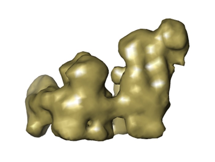
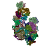
| Title | Three-dimensional structure of bovine respirasome by single particle cryo-electron tomography | |||||||||
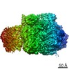
 Map data Map data | EM map of respirasome from bovine mitochondria | |||||||||
 Sample Sample |
| |||||||||
 Keywords Keywords | Oxidative phosphorylation / mitochondria | |||||||||
| Biological species |  | |||||||||
| Method | subtomogram averaging / cryo EM / negative staining / Resolution: 22.0 Å | |||||||||
 Authors Authors | Dudkina NV / Kudryashev M / Stahlberg H / Boekema EJ | |||||||||
 Citation Citation | Journal: Proc Natl Acad Sci U S A / Year: 2011 Title: Interaction of complexes I, III, and IV within the bovine respirasome by single particle cryoelectron tomography. Authors: Natalya V Dudkina / Mikhail Kudryashev / Henning Stahlberg / Egbert J Boekema /  Abstract: The respirasome is a multisubunit supercomplex of the respiratory chain in mitochondria. Here we report the 3D reconstruction of the bovine heart respirasome, composed of dimeric complex III and ...The respirasome is a multisubunit supercomplex of the respiratory chain in mitochondria. Here we report the 3D reconstruction of the bovine heart respirasome, composed of dimeric complex III and single copies of complex I and IV, at about 2.2-nm resolution, determined by cryoelectron tomography and subvolume averaging. Fitting of X-ray structures of single complexes I, III(2), and IV with high fidelity allows interpretation of the model at the level of secondary structures and shows how the individual complexes interact within the respirasome. Surprisingly, the distance between cytochrome c binding sites of complexes III(2) and IV is about 10 nm. Modeling indicates a loose interaction between the three complexes and provides evidence that lipids are gluing them at the interfaces. | |||||||||
| History |
|
- Structure visualization
| Movie |
 Movie viewer Movie viewer |
|---|---|
| Structure viewer | EM map: SurfViewMolmilJmol/JSmol |
| Supplemental images |
- Downloads & links
-EMDB archive
| Map data | emd_5319.map.gz | 7.4 MB | EMDB map data format | |
|---|---|---|---|---|
| Header (meta data) | emd-5319-v30.xmlemd-5319.xml | 13.4 KB 13.4 KB | Display Display | EMDB header |
| Images | emd_5319_1.tif | 210.2 KB | ||
| Archive directory |  http://ftp.pdbj.org/pub/emdb/structures/EMD-5319ftp://ftp.pdbj.org/pub/emdb/structures/EMD-5319 http://ftp.pdbj.org/pub/emdb/structures/EMD-5319ftp://ftp.pdbj.org/pub/emdb/structures/EMD-5319 | HTTPS FTP |
-Related structure data
| Similar structure data |
|---|


-Links
| EMDB pages | EMDB (EBI/PDBe) / EMDataResource |
|---|
-Map
| File | Download / File: emd_5319.map.gz / Format: CCP4 / Size: 7.8 MB / Type: IMAGE STORED AS FLOATING POINT NUMBER (4 BYTES) | ||||||||||||||||||||||||||||||||||||||||||||||||||||||||||||||||||||
|---|---|---|---|---|---|---|---|---|---|---|---|---|---|---|---|---|---|---|---|---|---|---|---|---|---|---|---|---|---|---|---|---|---|---|---|---|---|---|---|---|---|---|---|---|---|---|---|---|---|---|---|---|---|---|---|---|---|---|---|---|---|---|---|---|---|---|---|---|---|
| Annotation | EM map of respirasome from bovine mitochondria | ||||||||||||||||||||||||||||||||||||||||||||||||||||||||||||||||||||
| Projections & slices | Image control
Images are generated by Spider. | ||||||||||||||||||||||||||||||||||||||||||||||||||||||||||||||||||||
| Voxel size | X=Y=Z: 3.8 Å | ||||||||||||||||||||||||||||||||||||||||||||||||||||||||||||||||||||
| Density |
| ||||||||||||||||||||||||||||||||||||||||||||||||||||||||||||||||||||
| Symmetry | Space group: 1 | ||||||||||||||||||||||||||||||||||||||||||||||||||||||||||||||||||||
| Details | EMDB XML:
CCP4 map header:
| ||||||||||||||||||||||||||||||||||||||||||||||||||||||||||||||||||||
 Z (Sec.)
 Y (Row.)
 X (Col.)


-Supplemental data
- Sample components
-Entire : Respirasome from bovine mitochondria
| Entire | Name: Respirasome from bovine mitochondria |
|---|---|
| Components |
|
-Supramolecule #1000: Respirasome from bovine mitochondria
| Supramolecule | Name: Respirasome from bovine mitochondria / type: sample / ID: 1000 Details: The sample was thawed from storage at -80 degrees Celcius before being loaded onto the grid. Oligomeric state: One monomer of NADH dehydrogenase binds to one dimer of cytochrome bc1 and one monomer of cytochrome c oxidase Number unique components: 1 |
|---|---|
| Molecular weight | Experimental: 1.7 MDa / Theoretical: 1.7 MDa / Method: blue-native gel electrophoresis |
-Macromolecule #1: respirasome
| Macromolecule | Name: respirasome / type: protein_or_peptide / ID: 1 / Name.synonym: respirasome / Details: Solubilized with digitonin / Number of copies: 1 / Oligomeric state: monomer / Recombinant expression: No / Database: NCBI |
|---|---|
| Source (natural) | Organism: |
| Molecular weight | Experimental: 1.7 MDa / Theoretical: 1.7 MDa |
-Experimental details
-Structure determination
| Method | negative staining, cryo EM |
|---|---|
 Processing Processing | subtomogram averaging |
| Aggregation state | particle |
-Sample preparation
| Concentration | 0.5 mg/mL |
|---|---|
| Buffer | pH: 7 Details: 25 mM Tricine, 7.5 mM Bis-Tris, 0.1 mM PMSF, 0.01% digitonin |
| Staining | Type: NEGATIVE Details: 2.5 microliters were mixed with 10-nm gold particles as fiducial markers and applied on glow-discharged 200 mesh Quantifoil support grids with an additional carbon support film. Grids were ...Details: 2.5 microliters were mixed with 10-nm gold particles as fiducial markers and applied on glow-discharged 200 mesh Quantifoil support grids with an additional carbon support film. Grids were blotted at 100% humidity for 4-6 seconds at a blot offset (the longitudinal grid positioning) setting of -3.5, using a Vitrobot Mk3 (FEI). |
| Grid | Details: 200 mesh copper Quantifoil grids with an additional carbon support film |
| Vitrification | Cryogen name: ETHANE / Chamber humidity: 100 % / Chamber temperature: 89 K / Instrument: FEI VITROBOT MARK III / Details: Vitrification instrument: Vitrobot Mk3 (FEI) / Method: Blot for 4-6 seconds before plunging |
- Electron microscopy
| Microscope | FEI POLARA 300 |
|---|---|
| Temperature | Min: 15 K / Average: 77 K |
| Alignment procedure | Legacy - Astigmatism: objective lens astigmatism was corrected at 34,000 times magnification Legacy - Electron beam tilt params: 0 |
| Specialist optics | Energy filter - Name: FEI / Energy filter - Lower energy threshold: 0.0 eV / Energy filter - Upper energy threshold: 20.0 eV |
| Date | Mar 1, 2010 |
| Image recording | Category: CCD / Film or detector model: GENERIC GATAN (2k x 2k) / Digitization - Sampling interval: 0.38 µm / Number real images: 21 / Average electron dose: 80 e/Å2 |
| Electron beam | Acceleration voltage: 200 kV / Electron source:  FIELD EMISSION GUN FIELD EMISSION GUN |
| Electron optics | Illumination mode: SPOT SCAN / Imaging mode: BRIGHT FIELD / Cs: 2.0 mm / Nominal defocus max: 2.0 µm / Nominal defocus min: 2.0 µm / Nominal magnification: 34000 |
| Sample stage | Specimen holder: Multispecimen holder / Specimen holder model: SIDE ENTRY, EUCENTRIC / Tilt series - Axis1 - Min angle: -70 ° / Tilt series - Axis1 - Max angle: 70 ° |
| Experimental equipment |  Model: Tecnai Polara / Image courtesy: FEI Company |
-Image processing
| Details | The particles were selected from the tomograms by cross-correlation to an artificial globular reference followed by manual selection of particle volumes. Average number of projections used in the 3D reconstructions: 2466. Average number of class averages: 1. |
|---|---|
| Final reconstruction | Algorithm: OTHER / Resolution.type: BY AUTHOR / Resolution: 22.0 Å / Resolution method: OTHER / Software - Name: IMOD, XMIPP, AV3 |
-Atomic model buiding 1
| Initial model | PDB ID: |
|---|---|
| Software | Name: UCSF Chimera |
| Details | Protocol: Rigid Body |
| Refinement | Space: REAL / Protocol: RIGID BODY FIT / Target criteria: cross correlation |

-Atomic model buiding 2
| Initial model | PDB ID: |
|---|---|
| Software | Name: UCSF Chimera |
| Details | Protocol: Rigid Body |
| Refinement | Space: REAL / Protocol: RIGID BODY FIT / Target criteria: cross correlation |

-Atomic model buiding 3
| Initial model | PDB ID: |
|---|---|
| Software | Name: UCSF Chimera |
| Details | Protocol: Rigid Body |
| Refinement | Space: REAL / Protocol: RIGID BODY FIT / Target criteria: cross correlation |

