 Movie
Movie Controller

[English] 日本語
 Yorodumi
- PDB-3m9s: Crystal structure of respiratory complex I from Thermus thermophilus -
+ Open data

- Basic information
| Entry | Database: PDB / ID: 3m9s | ||||||
|---|---|---|---|---|---|---|---|
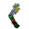
| Title | Crystal structure of respiratory complex I from Thermus thermophilus | ||||||
 Components Components |
| ||||||
 Keywords Keywords | OXIDOREDUCTASE / MEMBRANE PROTEIN / COMPLEX I / ELECTRON TRANSPORT / RESPIRATORY CHAIN | ||||||
| Function / homology |  Function and homology information Function and homology informationNADH dehydrogenase (quinone) (non-electrogenic) activity / Translocases; Catalysing the translocation of protons; Linked to oxidoreductase reactions / molybdopterin cofactor binding / iron-sulfur cluster assembly / electron transport coupled proton transport / NADH dehydrogenase activity / oxidoreductase activity, acting on NAD(P)H / respiratory chain complex I / NADH dehydrogenase (ubiquinone) activity / quinone binding ...NADH dehydrogenase (quinone) (non-electrogenic) activity / Translocases; Catalysing the translocation of protons; Linked to oxidoreductase reactions / molybdopterin cofactor binding / iron-sulfur cluster assembly / electron transport coupled proton transport / NADH dehydrogenase activity / oxidoreductase activity, acting on NAD(P)H / respiratory chain complex I / NADH dehydrogenase (ubiquinone) activity / quinone binding / ATP synthesis coupled electron transport / ferric iron binding / aerobic respiration / 2 iron, 2 sulfur cluster binding / NAD binding / FMN binding / 4 iron, 4 sulfur cluster binding / iron ion binding / metal ion binding / plasma membrane Similarity search - Function | ||||||
| Biological species |   Thermus thermophilus (bacteria) Thermus thermophilus (bacteria) | ||||||
| Method |  X-RAY DIFFRACTION / SYNCHROTRON / MOLECULAR REPLACEMENT / Resolution: 4.5 Å X-RAY DIFFRACTION / SYNCHROTRON / MOLECULAR REPLACEMENT / Resolution: 4.5 Å | ||||||
 Authors Authors | Efremov, R.G. / Baradaran, R. / Sazanov, L.A. | ||||||
 Citation Citation | Journal: Nature / Year: 2010 Title: The architecture of respiratory complex I Authors: Efremov, R.G. / Baradaran, R. / Sazanov, L.A. | ||||||
| History |
| ||||||
| Remark 650 | HELIX DETERMINATION METHOD: AUTHOR DETERMINED | ||||||
| Remark 700 | SHEET DETERMINATION METHOD: AUTHOR DETERMINED |
- Structure visualization
| Structure viewer | Molecule: MolmilJmol/JSmol |
|---|
- Downloads & links
-Download
| PDBx/mmCIF format | 3m9s.cif.gz | 1 MB | Display | PDBx/mmCIF format |
|---|---|---|---|---|
| PDB format | pdb3m9s.ent.gz | 810.2 KB | Display | PDB format |
| PDBx/mmJSON format | 3m9s.json.gz | Tree view | PDBx/mmJSON format | |
| Others |  Other downloads Other downloads |
-Validation report
| Arichive directory | https://data.pdbj.org/pub/pdb/validation_reports/m9/3m9sftp://data.pdbj.org/pub/pdb/validation_reports/m9/3m9s | HTTPS FTP |
|---|
-Related structure data
| Related structure data |  3m9cSC  3i9vS S: Starting model for refinement C: citing same article ( |
|---|---|
| Similar structure data |


-Links
 PDBj
PDBj


- Assembly
| Deposited unit | 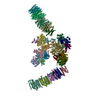
| ||||||||
|---|---|---|---|---|---|---|---|---|---|
| 1 | 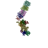
| ||||||||
| 2 | 
| ||||||||
| Unit cell |
| ||||||||
| Details | The biological assembly (complex I) consists of chains 1,2,3,4,5,6,7,8,9,L,M,N,R and H. / The identical biological assembly in ASU consists of chains A,B,C,D,E,F,J,I,G,O,P,Q,S and T. |
-Components
-NADH-quinone oxidoreductase subunit ... , 12 types, 24 molecules 1A2B3C4D5E6F9G7JLOMPNQHT
| #1: Protein | Mass: 48693.715 Da / Num. of mol.: 2 / Source method: isolated from a natural source / Source: (natural) Thermus thermophilus (bacteria) / Strain: HB8 / References: UniProt: Q56222, NADH dehydrogenase (quinone)#2: Protein | Mass: 20309.162 Da / Num. of mol.: 2 / Source method: isolated from a natural source / Source: (natural) Thermus thermophilus (bacteria) / Strain: HB8 / References: UniProt: Q56221, NADH dehydrogenase (quinone)#3: Protein | Mass: 86656.203 Da / Num. of mol.: 2 / Source method: isolated from a natural source / Source: (natural) Thermus thermophilus (bacteria) / Strain: HB8 / References: UniProt: Q56223, NADH dehydrogenase (quinone)#4: Protein | Mass: 46428.027 Da / Num. of mol.: 2 / Source method: isolated from a natural source / Source: (natural) Thermus thermophilus (bacteria) / Strain: HB8 / References: UniProt: Q56220, NADH dehydrogenase (quinone)#5: Protein | Mass: 23893.254 Da / Num. of mol.: 2 / Source method: isolated from a natural source / Source: (natural) Thermus thermophilus (bacteria) / Strain: HB8 / References: UniProt: Q56219, NADH dehydrogenase (quinone)#6: Protein | Mass: 20262.564 Da / Num. of mol.: 2 / Source method: isolated from a natural source / Source: (natural) Thermus thermophilus (bacteria) / Strain: HB8 / References: UniProt: Q56218, NADH dehydrogenase (quinone)#7: Protein | Mass: 20106.309 Da / Num. of mol.: 2 / Source method: isolated from a natural source / Source: (natural) Thermus thermophilus (bacteria) / Strain: HB8 / References: UniProt: Q56224, NADH dehydrogenase (quinone)#8: Protein | Mass: 14812.074 Da / Num. of mol.: 2 / Source method: isolated from a natural source / Source: (natural) Thermus thermophilus (bacteria) / Strain: HB8 / References: UniProt: Q5SKZ7, NADH dehydrogenase (quinone)#9: Protein | Mass: 39932.211 Da / Num. of mol.: 2 / Source method: isolated from a natural source / Source: (natural) Thermus thermophilus (bacteria) / Strain: HB8 / References: NADH dehydrogenase (quinone)#10: Protein | Mass: 33379.059 Da / Num. of mol.: 2 / Source method: isolated from a natural source / Source: (natural) Thermus thermophilus (bacteria) / Strain: HB8 / References: NADH:ubiquinone reductase (H+-translocating)#11: Protein | Mass: 32272.680 Da / Num. of mol.: 2 / Source method: isolated from a natural source / Source: (natural) Thermus thermophilus (bacteria) / Strain: HB8 / References: NADH:ubiquinone reductase (H+-translocating)#13: Protein | Mass: 15421.980 Da / Num. of mol.: 2 / Source method: isolated from a natural source / Source: (natural) Thermus thermophilus (bacteria) / Strain: HB8 / References: NADH:ubiquinone reductase (H+-translocating) |
|---|
-Protein , 1 types, 2 molecules RS
| #12: Protein | Mass: 23336.646 Da / Num. of mol.: 2 / Source method: isolated from a natural source / Source: (natural) Thermus thermophilus (bacteria) / Strain: HB8 / References: NADH:ubiquinone reductase (H+-translocating) |
|---|
-Non-polymers , 3 types, 20 molecules 


| #14: Chemical | ChemComp-SF4 /  Mass: 351.640 Da / Num. of mol.: 14 / Source method: obtained synthetically / Formula: Fe4S4 Mass: 351.640 Da / Num. of mol.: 14 / Source method: obtained synthetically / Formula: Fe4S4#15: Chemical |  Mass: 456.344 Da / Num. of mol.: 2 / Source method: obtained synthetically / Formula: C17H21N4O9P Mass: 456.344 Da / Num. of mol.: 2 / Source method: obtained synthetically / Formula: C17H21N4O9P#16: Chemical | ChemComp-FES /  Mass: 175.820 Da / Num. of mol.: 4 / Source method: obtained synthetically / Formula: Fe2S2 Mass: 175.820 Da / Num. of mol.: 4 / Source method: obtained synthetically / Formula: Fe2S2 |
|---|
-Details
| Compound details | THE LONG C-N BONDS ARE BECAUSE OF THE LOOPS NOT BEING MODELED. |
|---|---|
| Has protein modification | Y |
| Sequence details | THE ACTUAL SEQUENCE FOR CHAIN L,O: MALLGTILLPLLGFALLGLFGKRMREPLPGVLASGLVLASFLLGAGLLLSGGARFQAEWL ...THE ACTUAL SEQUENCE FOR CHAIN L,O: MALLGTILLP |
-Experimental details
-Experiment
| Experiment | Method: X-RAY DIFFRACTION / Number of used crystals: 1 |
|---|
- Sample preparation
| Crystal | Density Matthews: 4.91 Å3/Da / Density % sol: 68 % |
|---|---|
| Crystal grow | Temperature: 295 K / Method: vapor diffusion, sitting drop / pH: 6 Details: 16% PEG 4000, 100 mM Bis-Tris, 100 mM KCl, 100 mM glutaric acid and 2.04 mM n-octyl- -maltoside fluorinated, pH 6.0, VAPOR DIFFUSION, SITTING DROP, temperature 295K |
-Data collection
| Diffraction | Mean temperature: 100 K |
|---|---|
| Diffraction source | Source: SYNCHROTRON / Site: SLS  / Beamline: X06SA / Wavelength: 1 Å / Beamline: X06SA / Wavelength: 1 Å |
| Detector | Type: MARMOSAIC 225 mm CCD / Detector: CCD / Date: Jul 19, 2008 / Details: Mirrors |
| Radiation | Monochromator: Si(111) / Protocol: SINGLE WAVELENGTH / Monochromatic (M) / Laue (L): M / Scattering type: x-ray |
| Radiation wavelength | Wavelength: 1 Å / Relative weight: 1 |
| Reflection | Resolution: 4.5→30 Å / Num. all: 72549 / Num. obs: 72549 / % possible obs: 74.9 % / Observed criterion σ(F): 0 / Observed criterion σ(I): 0 / Redundancy: 1.8 % / Biso Wilson estimate: 132.9 Å2 / Rmerge(I) obs: 0.252 / Net I/σ(I): 2.5 |
| Reflection shell | Resolution: 4.5→4.74 Å / Redundancy: 1.6 % / Rmerge(I) obs: 0.628 / Mean I/σ(I) obs: 1 / Num. unique all: 9162 / % possible all: 65.1 |
- Processing
| Software |
| ||||||||||||
|---|---|---|---|---|---|---|---|---|---|---|---|---|---|
| Refinement | Method to determine structure: MOLECULAR REPLACEMENT Starting model: PDB ENTRIES 3I9V AND 3M9C Resolution: 4.5→30 Å / Num. reflection all: 72549 / Num. reflection obs: 72549 Details: The structure is not refined and consists of the atomic molecular replacement model of the hydrophilic domain and the backbone alpha-helical model of the membrane domain. Reflection data was ...Details: The structure is not refined and consists of the atomic molecular replacement model of the hydrophilic domain and the backbone alpha-helical model of the membrane domain. Reflection data was anisotropically scaled, for optimal electron density calculation. The directionality of the helices in the membrane domain is not known. The crystal is twinned (twin law "-h, -k, h+l"), with twin fraction 0.46. | ||||||||||||
| Refinement step | Cycle: LAST / Resolution: 4.5→30 Å
|
