 Movie
Movie Controller

[English] 日本語
 Yorodumi
- PDB-3m9c: Crystal structure of the membrane domain of respiratory complex I... -
+ Open data

- Basic information
| Entry | Database: PDB / ID: 3m9c | ||||||
|---|---|---|---|---|---|---|---|
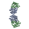
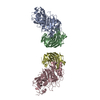
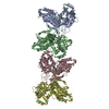
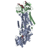
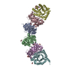
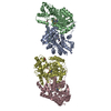
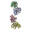
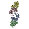
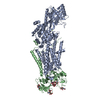
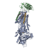
| Title | Crystal structure of the membrane domain of respiratory complex I from Escherichia coli | ||||||
 Components Components |
| ||||||
 Keywords Keywords | OXIDOREDUCTASE / MEMBRANE PROTEIN / COMPLEX I / ELECTRON TRANSPORT / RESPIRATORY CHAIN | ||||||
| Biological species |  | ||||||
| Method |  X-RAY DIFFRACTION / SYNCHROTRON / MIRAS / Resolution: 3.9 Å X-RAY DIFFRACTION / SYNCHROTRON / MIRAS / Resolution: 3.9 Å | ||||||
 Authors Authors | Efremov, R.G. / Baradaran, R. / Sazanov, L.A. | ||||||
 Citation Citation | Journal: Nature / Year: 2010 Title: The architecture of respiratory complex I Authors: Efremov, R.G. / Baradaran, R. / Sazanov, L.A. | ||||||
| History |
| ||||||
| Remark 650 | HELIX DETERMINATION METHOD: AUTHOR DETERMINED |
- Structure visualization
| Structure viewer | Molecule:  MolmilJmol/JSmol MolmilJmol/JSmol |
|---|
- Downloads & links
-Download
| PDBx/mmCIF format | 3m9c.cif.gz | 110.8 KB | Display | PDBx/mmCIF format |
|---|---|---|---|---|
| PDB format | pdb3m9c.ent.gz | 81.6 KB | Display | PDB format |
| PDBx/mmJSON format | 3m9c.json.gz | Tree view | PDBx/mmJSON format | |
| Others |  Other downloads Other downloads |
-Validation report
| Arichive directory | https://data.pdbj.org/pub/pdb/validation_reports/m9/3m9cftp://data.pdbj.org/pub/pdb/validation_reports/m9/3m9c | HTTPS FTP |
|---|
-Related structure data

-Links
 PDBj
PDBj
- Assembly
| Deposited unit | 
| ||||||||
|---|---|---|---|---|---|---|---|---|---|
| 1 |
| ||||||||
| Unit cell |
|
-Components
| #1: Protein | Mass: 40357.742 Da / Num. of mol.: 1 / Fragment: Membrane domain / Mutation: Subunit NuoH is missing / Source method: isolated from a natural source / Source: (natural) |
|---|---|
| #2: Protein | Mass: 33293.949 Da / Num. of mol.: 1 / Fragment: Membrane domain / Mutation: Subunit NuoH is missing / Source method: isolated from a natural source / Source: (natural) |
| #3: Protein | Mass: 32187.574 Da / Num. of mol.: 1 / Fragment: Membrane domain / Mutation: Subunit NuoH is missing / Source method: isolated from a natural source / Source: (natural) |
| #4: Protein | Mass: 23932.375 Da / Num. of mol.: 1 / Fragment: Membrane domain / Mutation: Subunit NuoH is missing / Source method: isolated from a natural source / Source: (natural) |
| Compound details | THE LONG C-N BONDS ARE BECAUSE OF THE LOOPS NOT BEING MODELED. |
| Sequence details | THE ACTUAL SEQUENCE FOR CHAIN L: MNMLALTIILPLIGFVLLAFSRGRWSENVSAIVGVGSVGLAALVTAFIGVDFFANGEQTYSQPLW ...THE ACTUAL SEQUENCE FOR CHAIN L: MNMLALTIIL |
-Experimental details
-Experiment
| Experiment | Method: X-RAY DIFFRACTION / Number of used crystals: 1 |
|---|
- Sample preparation
| Crystal | Density Matthews: 7.42 Å3/Da / Density % sol: 73 % |
|---|---|
| Crystal grow | Temperature: 283 K / Method: vapor diffusion, sitting drop / pH: 4.8 Details: 9% PEG4000, 0.1 M sodium acetate, 1.0 M sodium formate, pH 4.8, VAPOR DIFFUSION, SITTING DROP, temperature 283K |
-Data collection
| Diffraction | Mean temperature: 100 K |
|---|---|
| Diffraction source | Source: SYNCHROTRON / Site: ESRF  / Beamline: ID23-2 / Wavelength: 0.873 Å / Beamline: ID23-2 / Wavelength: 0.873 Å |
| Detector | Type: MARMOSAIC 225 mm CCD / Detector: CCD / Date: Feb 21, 2009 / Details: Mirrors |
| Radiation | Monochromator: Si 111 / Protocol: SINGLE WAVELENGTH / Monochromatic (M) / Laue (L): M / Scattering type: x-ray |
| Radiation wavelength | Wavelength: 0.873 Å / Relative weight: 1 |
| Reflection | Resolution: 3.9→30 Å / Num. all: 34983 / Num. obs: 34983 / % possible obs: 97.6 % / Observed criterion σ(F): 0 / Observed criterion σ(I): 0 / Redundancy: 3.3 % / Biso Wilson estimate: 82.3 Å2 / Rmerge(I) obs: 0.205 / Net I/σ(I): 4 |
| Reflection shell | Resolution: 3.9→4.11 Å / Redundancy: 3.2 % / Rmerge(I) obs: 0.846 / Mean I/σ(I) obs: 1.5 / Num. unique all: 5022 / % possible all: 97.4 |
- Processing
| Software |
| ||||||||||||||
|---|---|---|---|---|---|---|---|---|---|---|---|---|---|---|---|
| Refinement | Method to determine structure: MIRAS / Resolution: 3.9→30 Å / Num. reflection all: 34983 / Num. reflection obs: 34983 Details: This is not refined backbone model. The directionality of alpha-helices is not known. For the optimal calculation of electron density the data should be anisotropically scaled. | ||||||||||||||
| Refinement step | Cycle: LAST / Resolution: 3.9→30 Å
|
