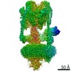
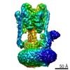
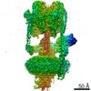
Journal: DNA Repair (Amst) / Year: 2020 Title: A combined structural and biochemical approach reveals translocation and stalling of UvrB on the DNA lesion as a mechanism of damage verification in bacterial nucleotide excision repair. Authors: Marcin Jaciuk / Paolo Swuec / Vineet Gaur / Joanna M Kasprzak / Ludovic Renault / Mateusz Dobrychłop / Shivlee Nirwal / Janusz M Bujnicki / Alessandro Costa / Marcin Nowotny / Abstract: Nucleotide excision repair (NER) is a DNA repair pathway present in all domains of life. In bacteria, UvrA protein localizes the DNA lesion, followed by verification by UvrB helicase and excision by ...Nucleotide excision repair (NER) is a DNA repair pathway present in all domains of life. In bacteria, UvrA protein localizes the DNA lesion, followed by verification by UvrB helicase and excision by UvrC double nuclease. UvrA senses deformations and flexibility of the DNA duplex without precisely localizing the lesion in the damaged strand, an element essential for proper NER. Using a combination of techniques, we elucidate the mechanism of the damage verification step in bacterial NER. UvrA dimer recruits two UvrB molecules to its two sides. Each of the two UvrB molecules clamps a different DNA strand using its β-hairpin element. Both UvrB molecules then translocate to the lesion, and UvrA dissociates. The UvrB molecule that clamps the damaged strand gets stalled at the lesion to recruit UvrC. This mechanism allows UvrB to verify the DNA damage and identify its precise location triggering subsequent steps in the NER pathway.
History
Deposition
May 9, 2019
-
Header (metadata) release
May 22, 2019
-
Map release
May 22, 2019
-
Update
Dec 4, 2019
-
Current status
Dec 4, 2019
Processing site: PDBe / Status: Released
-
Structure visualization
Movie
Surface view with section colored by density value
In the structure databanks used in Yorodumi, some data are registered as the other names, "COVID-19 virus" and "2019-nCoV". Here are the details of the virus and the list of structure data.
Jan 31, 2019. EMDB accession codes are about to change! (news from PDBe EMDB page)
EMDB accession codes are about to change! (news from PDBe EMDB page)
The allocation of 4 digits for EMDB accession codes will soon come to an end. Whilst these codes will remain in use, new EMDB accession codes will include an additional digit and will expand incrementally as the available range of codes is exhausted. The current 4-digit format prefixed with “EMD-” (i.e. EMD-XXXX) will advance to a 5-digit format (i.e. EMD-XXXXX), and so on. It is currently estimated that the 4-digit codes will be depleted around Spring 2019, at which point the 5-digit format will come into force.
The EM Navigator/Yorodumi systems omit the EMD- prefix.
Related info.:Q: What is EMD? / ID/Accession-code notation in Yorodumi/EM Navigator
Yorodumi is a browser for structure data from EMDB, PDB, SASBDB, etc.
This page is also the successor to EM Navigator detail page, and also detail information page/front-end page for Omokage search.
The word "yorodu" (or yorozu) is an old Japanese word meaning "ten thousand". "mi" (miru) is to see.
Related info.:EMDB / PDB / SASBDB / Comparison of 3 databanks / Yorodumi Search / Aug 31, 2016. New EM Navigator & Yorodumi / Yorodumi Papers / Jmol/JSmol / Function and homology information / Changes in new EM Navigator and Yorodumi
 Movie
Movie Controller

 Yorodumi
 Open data
 Basic information
 Map data
 Sample
 Thermotoga maritima (bacteria)
 Authors
 Poland,
  United Kingdom, 4 items
  Citation
 Structure visualization
 Movie viewer
 UCSF Chimera
 Downloads & links
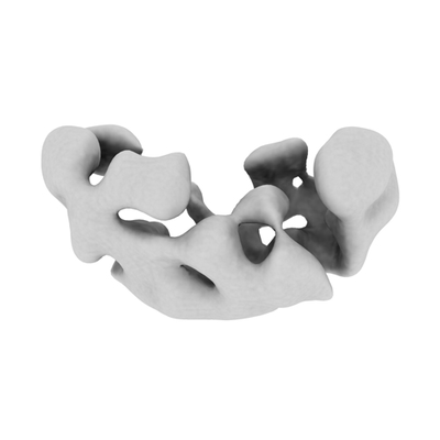 emd_4958.png
 http://ftp.pdbj.org/pub/emdb/structures/EMD-4958
 Z (Sec.)
 Y (Row.)
 X (Col.)


 Sample components
 Processing
 Electron microscopy