 ムービー
ムービー コントローラー


+ データを開く

- 基本情報
基本情報
| 登録情報 |  | ||||||||||||
|---|---|---|---|---|---|---|---|---|---|---|---|---|---|
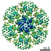
| タイトル | HIV-2 CA hexamer; assembled via liposome templating | ||||||||||||
 マップデータ マップデータ | EM map of the HIV-2 CA hexamer surrounded by CA hexamers assembled via templating on functionalized liposomes. | ||||||||||||
 試料 試料 |
| ||||||||||||
 キーワード キーワード | HIV-2 / Capsid / IP6 / VIRAL PROTEIN | ||||||||||||
| 機能・相同性 |  機能・相同性情報 機能・相同性情報HIV-2 retropepsin / retroviral ribonuclease H / exoribonuclease H / exoribonuclease H activity / DNA integration / viral genome integration into host DNA / establishment of integrated proviral latency / RNA-directed DNA polymerase / RNA stem-loop binding / viral penetration into host nucleus ...HIV-2 retropepsin / retroviral ribonuclease H / exoribonuclease H / exoribonuclease H activity / DNA integration / viral genome integration into host DNA / establishment of integrated proviral latency / RNA-directed DNA polymerase / RNA stem-loop binding / viral penetration into host nucleus / host multivesicular body / RNA-directed DNA polymerase activity / RNA-DNA hybrid ribonuclease activity / 転移酵素; リンを含む基を移すもの; 核酸を移すもの / host cell / viral nucleocapsid / DNA recombination / DNA-directed DNA polymerase / aspartic-type endopeptidase activity / 加水分解酵素; エステル加水分解酵素 / DNA-directed DNA polymerase activity / symbiont-mediated suppression of host gene expression / viral translational frameshifting / symbiont entry into host cell / lipid binding / host cell plasma membrane / host cell nucleus / virion membrane / structural molecule activity / proteolysis / DNA binding / zinc ion binding 類似検索 - 分子機能 | ||||||||||||
| 生物種 |  Human immunodeficiency virus 2 (ヒト免疫不全ウイルス) Human immunodeficiency virus 2 (ヒト免疫不全ウイルス) | ||||||||||||
| 手法 | 単粒子再構成法 / クライオ電子顕微鏡法 / 解像度: 3.25 Å | ||||||||||||
 データ登録者 データ登録者 | Freniere C / Cook M / Xiong Y | ||||||||||||
| 資金援助 |  米国, 3件 米国, 3件
| ||||||||||||
 引用 引用 | ジャーナル: Cell Rep / 年: 2025 タイトル: Structural insights into HIV-2 CA lattice formation and FG-pocket binding revealed by single-particle cryo-EM. 著者: Matthew Cook / Christian Freniere / Chunxiang Wu / Faith Lozano / Yong Xiong / 要旨: One of the striking features of human immunodeficiency virus (HIV) is the capsid, a fullerene cone comprised of pleomorphic capsid protein (CA) that shields the viral genome and recruits cofactors. ...One of the striking features of human immunodeficiency virus (HIV) is the capsid, a fullerene cone comprised of pleomorphic capsid protein (CA) that shields the viral genome and recruits cofactors. Despite significant advances in understanding the mechanisms of HIV-1 CA assembly and host factor interactions, HIV-2 CA assembly remains poorly understood. By templating the assembly of HIV-2 CA on functionalized liposomes, we report high-resolution structures of the HIV-2 CA lattice, including both CA hexamers and pentamers, alone and with peptides of host phenylalanine-glycine (FG)-motif proteins Nup153 and CPSF6. While the overall fold and mode of FG-peptide binding is conserved with HIV-1, this study reveals distinctive features of the HIV-2 CA lattice, including differing structural character at regions of host factor interactions and divergence in the mechanism of formation of CA hexamers and pentamers. This study extends our understanding of HIV capsids and highlights an approach facilitating the study of lentiviral capsid biology. | ||||||||||||
| 履歴 |
|
- 構造の表示
構造の表示
| 添付画像 |
|---|

- ダウンロードとリンク
-EMDBアーカイブ
| マップデータ | emd_45758.map.gz | 126.8 MB | EMDBマップデータ形式 | |
|---|---|---|---|---|
| ヘッダ (付随情報) | emd-45758-v30.xmlemd-45758.xml | 22.3 KB 22.3 KB | 表示 表示 | EMDBヘッダ |
| FSC (解像度算出) | emd_45758_fsc.xml | 13.7 KB | 表示 | FSCデータファイル |
| 画像 |  emd_45758.png emd_45758.png | 273.9 KB | ||
| Filedesc metadata | emd-45758.cif.gz | 7.1 KB | ||
| その他 | emd_45758_half_map_1.map.gzemd_45758_half_map_2.map.gz | 251.6 MB 251.6 MB | ||
| アーカイブディレクトリ |  http://ftp.pdbj.org/pub/emdb/structures/EMD-45758ftp://ftp.pdbj.org/pub/emdb/structures/EMD-45758 http://ftp.pdbj.org/pub/emdb/structures/EMD-45758ftp://ftp.pdbj.org/pub/emdb/structures/EMD-45758 | HTTPS FTP |
-関連構造データ


-リンク
| EMDBのページ | EMDB (EBI/PDBe) / EMDataResource |
|---|---|
| 「今月の分子」の関連する項目 |

-マップ
| ファイル | ダウンロード / ファイル: emd_45758.map.gz / 形式: CCP4 / 大きさ: 274.6 MB / タイプ: IMAGE STORED AS FLOATING POINT NUMBER (4 BYTES) | ||||||||||||||||||||||||||||||||||||
|---|---|---|---|---|---|---|---|---|---|---|---|---|---|---|---|---|---|---|---|---|---|---|---|---|---|---|---|---|---|---|---|---|---|---|---|---|---|
| 注釈 | EM map of the HIV-2 CA hexamer surrounded by CA hexamers assembled via templating on functionalized liposomes. | ||||||||||||||||||||||||||||||||||||
| 投影像・断面図 | 画像のコントロール
画像は Spider により作成 | ||||||||||||||||||||||||||||||||||||
| ボクセルのサイズ | X=Y=Z: 1.086 Å | ||||||||||||||||||||||||||||||||||||
| 密度 |
| ||||||||||||||||||||||||||||||||||||
| 対称性 | 空間群: 1 | ||||||||||||||||||||||||||||||||||||
| 詳細 | EMDB XML:
|
 Z (Sec.)
 Y (Row.)
 X (Col.)


-添付データ
-ハーフマップ: EM half map B of the HIV-2 CA hexamer surrounded by CA hexamers.
| ファイル | emd_45758_half_map_1.map | ||||||||||||
|---|---|---|---|---|---|---|---|---|---|---|---|---|---|
| 注釈 | EM half map B of the HIV-2 CA hexamer surrounded by CA hexamers. | ||||||||||||
| 投影像・断面図 |
| ||||||||||||
| 密度ヒストグラム |


-ハーフマップ: EM half map A of the HIV-2 CA hexamer surrounded by CA hexamers.
| ファイル | emd_45758_half_map_2.map | ||||||||||||
|---|---|---|---|---|---|---|---|---|---|---|---|---|---|
| 注釈 | EM half map A of the HIV-2 CA hexamer surrounded by CA hexamers. | ||||||||||||
| 投影像・断面図 |
| ||||||||||||
| 密度ヒストグラム |


- 試料の構成要素
-全体 : HIV-2 capsid protein assembled into a lattice via liposome templating.
| 全体 | 名称: HIV-2 capsid protein assembled into a lattice via liposome templating. |
|---|---|
| 要素 |
|
-超分子 #1: HIV-2 capsid protein assembled into a lattice via liposome templating.
| 超分子 | 名称: HIV-2 capsid protein assembled into a lattice via liposome templating. タイプ: complex / ID: 1 / 親要素: 0 / 含まれる分子: #1 詳細: C-terminally hexahistidine tagged HIV-2 CA associated with a liposome decorated with NiNTA headgroups which results in the assembly of a lattice of CA. |
|---|---|
| 由来(天然) | 生物種: Human immunodeficiency virus 2 (ヒト免疫不全ウイルス) 株: GL-AN |
| 分子量 | 理論値: 26.9 KDa |
-分子 #1: Capsid protein p24
| 分子 | 名称: Capsid protein p24 / タイプ: protein_or_peptide / ID: 1 詳細: HIV-2 GL-AN capsid protein with C-terminal Gly-Ser-Ser linker to hexahistidine tag following proteolytic cleavage of the N-terminal Met. コピー数: 3 / 光学異性体: LEVO |
|---|---|
| 由来(天然) | 生物種: Human immunodeficiency virus 2 (ヒト免疫不全ウイルス) 株: GL-AN |
| 分子量 | 理論値: 26.80949 KDa |
| 組換発現 | 生物種:  |
| 配列 | 文字列: PVQQTGGGNY IHVPLSPRTL NAWVKLVEDK KFGAEVVPGF QALSEGCTPY DINQMLNCVG DHQAAMQIIR EIINDEAADW DAQHPIPGP LPAGQLRDPR GSDIAGTTST VEEQIQWMYR PQNPVPVGNI YRRWIQIGLQ KCVRMYNPTN ILDVKQGPKE P FQSYVDRF ...文字列: PVQQTGGGNY IHVPLSPRTL NAWVKLVEDK KFGAEVVPGF QALSEGCTPY DINQMLNCVG DHQAAMQIIR EIINDEAADW DAQHPIPGP LPAGQLRDPR GSDIAGTTST VEEQIQWMYR PQNPVPVGNI YRRWIQIGLQ KCVRMYNPTN ILDVKQGPKE P FQSYVDRF YKSLRAEQTD PAVKNWMTQT LLIQNANPDC KLVLKGLGMN PTLEEMLTAC QGVGGPGQKA RLMGSSHHHH HH UniProtKB: Gag-Pol polyprotein |
-分子 #2: INOSITOL HEXAKISPHOSPHATE
| 分子 | 名称: INOSITOL HEXAKISPHOSPHATE / タイプ: ligand / ID: 2 / コピー数: 2 / 式: IHP |
|---|---|
| 分子量 | 理論値: 660.035 Da |
| Chemical component information |  ChemComp-IHP: |
-実験情報
-構造解析
| 手法 | クライオ電子顕微鏡法 |
|---|---|
 解析 解析 | 単粒子再構成法 |
| 試料の集合状態 | particle |
-試料調製
| 濃度 | 10.7 mg/mL | |||||||||||||||
|---|---|---|---|---|---|---|---|---|---|---|---|---|---|---|---|---|
| 緩衝液 | pH: 7 構成要素:
詳細: The mixed buffer of storage buffer for the protein and lipid components with IP6 supplemented. | |||||||||||||||
| グリッド | モデル: Quantifoil R2/1 / 材質: COPPER / メッシュ: 200 / 支持フィルム - 材質: CARBON / 支持フィルム - トポロジー: HOLEY ARRAY / 前処理 - タイプ: GLOW DISCHARGE / 前処理 - 時間: 45 sec. / 前処理 - 雰囲気: AIR / 前処理 - 気圧: 0.015 kPa / 詳細: 15 mA discharge current. | |||||||||||||||
| 凍結 | 凍結剤: ETHANE / チャンバー内湿度: 100 % / チャンバー内温度: 298 K / 装置: FEI VITROBOT MARK IV 詳細: Grids were dual-side blotted with blot force 0 for 5.5 sec before plunge freezing in liquid ethane.. | |||||||||||||||
| 詳細 | Sample was prepared with 400 uM HIV-2 CA-6xHis protein, 5.9 mM lipid mix (described in publication), and 4 mM IP6. Sample was well-distributed on the grid, mostly monodisperse. Perhaps slightly more particles on carbon versus in the hole. |
- 電子顕微鏡法
| 顕微鏡 | FEI TITAN KRIOS |
|---|---|
| 特殊光学系 | エネルギーフィルター - 名称: GIF Quantum LS / エネルギーフィルター - スリット幅: 20 eV |
| 撮影 | フィルム・検出器のモデル: GATAN K3 (6k x 4k) / 平均電子線量: 50.0 e/Å2 |
| 電子線 | 加速電圧: 300 kV / 電子線源:  FIELD EMISSION GUN FIELD EMISSION GUN |
| 電子光学系 | C2レンズ絞り径: 30.0 µm / 照射モード: FLOOD BEAM / 撮影モード: BRIGHT FIELD / Cs: 2.7 mm / 最大 デフォーカス(公称値): 2.0 µm / 最小 デフォーカス(公称値): 0.8 µm / 倍率(公称値): 81000 |
| 試料ステージ | 試料ホルダーモデル: FEI TITAN KRIOS AUTOGRID HOLDER ホルダー冷却材: NITROGEN |
| 実験機器 |  モデル: Titan Krios / 画像提供: FEI Company |
+画像解析

-原子モデル構築 1
| 初期モデル | PDB ID: Chain - Chain ID: A / Chain - Residue range: 1-223 / Chain - Source name: Other / Chain - Initial model type: experimental model / 詳細: NTDs matched well, but the CTD had to be realigned. |
|---|---|
| 詳細 | The HIV-2 CA pentamer chain derived from micelle-templated icosahedra described in this publication was used as an initial model for fitting. Flexible fitting was used to move the CTD into its proper location. |
| 精密化 | 空間: REAL / プロトコル: FLEXIBLE FIT 当てはまり具合の基準: Cross-correlation coefficient |
| 得られたモデル |  PDB-9cns: |
