 Movie
Movie Controller

[English] 日本語
 Yorodumi
- EMDB-45762: HIV-2 CA pentamer in the presence of Nup153 peptide but without b... -
+ Open data

- Basic information
| Entry |  | ||||||||||||
|---|---|---|---|---|---|---|---|---|---|---|---|---|---|
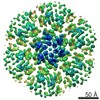
| Title | HIV-2 CA pentamer in the presence of Nup153 peptide but without binding; assembled via liposome templating | ||||||||||||
 Map data Map data | EM map of an HIV-2 CA pentamer assembled via templating on functionalized liposomes with Nup153 peptide introduced after, though no Nup153 peptide density is observed. | ||||||||||||
 Sample Sample |
| ||||||||||||
 Keywords Keywords | HIV-2 / Capsid / IP6 / Nup153 / VIRAL PROTEIN | ||||||||||||
| Biological species |  Human immunodeficiency virus 2 / Human immunodeficiency virus 2 /  Homo sapiens (human) Homo sapiens (human) | ||||||||||||
| Method | single particle reconstruction / cryo EM / Resolution: 2.98 Å | ||||||||||||
 Authors Authors | Freniere C / Cook M / Xiong Y | ||||||||||||
| Funding support |  United States, 3 items United States, 3 items
| ||||||||||||
 Citation Citation | Journal: Cell Rep / Year: 2025 Title: Structural insights into HIV-2 CA lattice formation and FG-pocket binding revealed by single-particle cryo-EM. Authors: Matthew Cook / Christian Freniere / Chunxiang Wu / Faith Lozano / Yong Xiong / Abstract: One of the striking features of human immunodeficiency virus (HIV) is the capsid, a fullerene cone comprised of pleomorphic capsid protein (CA) that shields the viral genome and recruits cofactors. ...One of the striking features of human immunodeficiency virus (HIV) is the capsid, a fullerene cone comprised of pleomorphic capsid protein (CA) that shields the viral genome and recruits cofactors. Despite significant advances in understanding the mechanisms of HIV-1 CA assembly and host factor interactions, HIV-2 CA assembly remains poorly understood. By templating the assembly of HIV-2 CA on functionalized liposomes, we report high-resolution structures of the HIV-2 CA lattice, including both CA hexamers and pentamers, alone and with peptides of host phenylalanine-glycine (FG)-motif proteins Nup153 and CPSF6. While the overall fold and mode of FG-peptide binding is conserved with HIV-1, this study reveals distinctive features of the HIV-2 CA lattice, including differing structural character at regions of host factor interactions and divergence in the mechanism of formation of CA hexamers and pentamers. This study extends our understanding of HIV capsids and highlights an approach facilitating the study of lentiviral capsid biology. | ||||||||||||
| History |
|
- Structure visualization
| Supplemental images |
|---|

- Downloads & links
-EMDB archive
| Map data | emd_45762.map.gz | 10.5 MB |  EMDB map data format EMDB map data format | |
|---|---|---|---|---|
| Header (meta data) | emd-45762-v30.xmlemd-45762.xml | 23 KB 23 KB | Display Display | EMDB header |
| FSC (resolution estimation) | emd_45762_fsc.xml | 5.9 KB | Display | FSC data file |
| Images |  emd_45762.png emd_45762.png | 247.1 KB | ||
| Filedesc metadata | emd-45762.cif.gz | 6.7 KB | ||
| Others | emd_45762_half_map_1.map.gzemd_45762_half_map_2.map.gz | 19.9 MB 19.9 MB | ||
| Archive directory |  http://ftp.pdbj.org/pub/emdb/structures/EMD-45762ftp://ftp.pdbj.org/pub/emdb/structures/EMD-45762 http://ftp.pdbj.org/pub/emdb/structures/EMD-45762ftp://ftp.pdbj.org/pub/emdb/structures/EMD-45762 | HTTPS FTP |
-Related structure data


-Links
| EMDB pages | EMDB (EBI/PDBe) / EMDataResource |
|---|
-Map
| File | Download / File: emd_45762.map.gz / Format: CCP4 / Size: 22.2 MB / Type: IMAGE STORED AS FLOATING POINT NUMBER (4 BYTES) | ||||||||||||||||||||||||||||||||||||
|---|---|---|---|---|---|---|---|---|---|---|---|---|---|---|---|---|---|---|---|---|---|---|---|---|---|---|---|---|---|---|---|---|---|---|---|---|---|
| Annotation | EM map of an HIV-2 CA pentamer assembled via templating on functionalized liposomes with Nup153 peptide introduced after, though no Nup153 peptide density is observed. | ||||||||||||||||||||||||||||||||||||
| Projections & slices | Image control
Images are generated by Spider. | ||||||||||||||||||||||||||||||||||||
| Voxel size | X=Y=Z: 1.068 Å | ||||||||||||||||||||||||||||||||||||
| Density |
| ||||||||||||||||||||||||||||||||||||
| Symmetry | Space group: 1 | ||||||||||||||||||||||||||||||||||||
| Details | EMDB XML:
|
 Z (Sec.)
 Y (Row.)
 X (Col.)


-Supplemental data
-Half map: EM half map A of an HIV-2 CA...
| File | emd_45762_half_map_1.map | ||||||||||||
|---|---|---|---|---|---|---|---|---|---|---|---|---|---|
| Annotation | EM half map A of an HIV-2 CA pentamer in the presence of Nup153 peptide, though density isn't apparent. | ||||||||||||
| Projections & Slices |
| ||||||||||||
| Density Histograms |


-Half map: EM half map B of an HIV-2 CA...
| File | emd_45762_half_map_2.map | ||||||||||||
|---|---|---|---|---|---|---|---|---|---|---|---|---|---|
| Annotation | EM half map B of an HIV-2 CA pentamer in the presence of Nup153 peptide, though density isn't apparent. | ||||||||||||
| Projections & Slices |
| ||||||||||||
| Density Histograms |


- Sample components
-Entire : HIV-2 capsid protein assembled into a lattice via liposome templa...
| Entire | Name: HIV-2 capsid protein assembled into a lattice via liposome templating and then Nup153 peptide is introduced. |
|---|---|
| Components |
|
-Supramolecule #1: HIV-2 capsid protein assembled into a lattice via liposome templa...
| Supramolecule | Name: HIV-2 capsid protein assembled into a lattice via liposome templating and then Nup153 peptide is introduced. type: complex / ID: 1 / Parent: 0 / Macromolecule list: all Details: C-terminally hexahistidine tagged HIV-2 CA associated with a liposome decorated with NiNTA headgroups which results in the assembly of a lattice of CA. Nup153 peptide is subsequently ...Details: C-terminally hexahistidine tagged HIV-2 CA associated with a liposome decorated with NiNTA headgroups which results in the assembly of a lattice of CA. Nup153 peptide is subsequently introduced and potential binding is allowed to equilibrate. |
|---|---|
| Molecular weight | Theoretical: 26.9 KDa |
-Supramolecule #2: HIV-2 capsid protein
| Supramolecule | Name: HIV-2 capsid protein / type: complex / ID: 2 / Parent: 1 / Macromolecule list: #1 |
|---|---|
| Source (natural) | Organism: Human immunodeficiency virus 2 / Strain: GL-AN |
-Supramolecule #3: Nup153 peptide
| Supramolecule | Name: Nup153 peptide / type: complex / ID: 3 / Parent: 1 / Macromolecule list: #2 |
|---|---|
| Source (natural) | Organism: Homo sapiens (human) / Synthetically produced: Yes |
-Macromolecule #1: HIV-2 capsid protein
| Macromolecule | Name: HIV-2 capsid protein / type: protein_or_peptide / ID: 1 Details: HIV-2 capsid protein with C-terminal Gly-Ser-Ser linker and hexahistidine tag following proteolytic processing of the N-terminal Met. Enantiomer: LEVO |
|---|---|
| Source (natural) | Organism: Human immunodeficiency virus 2 / Strain: GL-AN |
| Recombinant expression | Organism:  |
| Sequence | String: PVQQTGGGNY IHVPLSPRTL NAWVKLVEDK KFGAEVVPGF QALSEGCTPY DINQMLNCVG DHQAAMQIIR EIINDEAADW DAQHPIPGPL PAGQLRDPRG SDIAGTTSTV EEQIQWMYRP QNPVPVGNIY RRWIQIGLQK CVRMYNPTNI LDVKQGPKEP FQSYVDRFYK ...String: PVQQTGGGNY IHVPLSPRTL NAWVKLVEDK KFGAEVVPGF QALSEGCTPY DINQMLNCVG DHQAAMQIIR EIINDEAADW DAQHPIPGPL PAGQLRDPRG SDIAGTTSTV EEQIQWMYRP QNPVPVGNIY RRWIQIGLQK CVRMYNPTNI LDVKQGPKEP FQSYVDRFYK SLRAEQTDPA VKNWMTQTLL IQNANPDCKL VLKGLGMNPT LEEMLTACQG VGGPGQKARL MGSSHHHHHH |
-Macromolecule #2: Nup153
| Macromolecule | Name: Nup153 / type: protein_or_peptide / ID: 2 Details: Peptide of Nup153, residues 1411-1425 and 1464-1475 Enantiomer: LEVO |
|---|---|
| Source (natural) | Organism: Homo sapiens (human) |
| Sequence | String: PSGVFTFGAN SSTPAGRKIK TAVRRRK |
-Experimental details
-Structure determination
| Method | cryo EM |
|---|---|
 Processing Processing | single particle reconstruction |
| Aggregation state | particle |
-Sample preparation
| Concentration | 10.7 mg/mL | |||||||||||||||
|---|---|---|---|---|---|---|---|---|---|---|---|---|---|---|---|---|
| Buffer | pH: 7 Component:
Details: The mixed buffer of storage buffer for the protein and lipid components with IP6 supplemented. | |||||||||||||||
| Grid | Model: Quantifoil R2/1 / Material: COPPER / Mesh: 200 / Support film - Material: CARBON / Support film - topology: HOLEY ARRAY / Support film - Film thickness: 12 / Pretreatment - Type: GLOW DISCHARGE / Pretreatment - Time: 45 sec. / Pretreatment - Atmosphere: AIR / Pretreatment - Pressure: 0.015 kPa / Details: 15 mA discharge current. | |||||||||||||||
| Vitrification | Cryogen name: ETHANE / Chamber humidity: 100 % / Chamber temperature: 298 K / Instrument: FEI VITROBOT MARK IV Details: Grids were dual-side blotted with blot force 0 for 5.5 sec before plunge freezing in liquid ethane.. | |||||||||||||||
| Details | Sample was prepared with 400 uM HIV-2 CA-6xHis protein, 5.9 mM lipid mix (described in publication), and 4 mM IP6 final concentrations after subsequent addition. Nup153 peptide was then introduced to 400 uM final concentration. Sample was well-distributed on the grid, mostly monodisperse. Perhaps slightly more particles on carbon versus in the hole. |
- Electron microscopy
| Microscope | FEI TITAN KRIOS |
|---|---|
| Specialist optics | Energy filter - Name: GIF Quantum LS / Energy filter - Slit width: 20 eV |
| Image recording | Film or detector model: GATAN K3 (6k x 4k) / Average electron dose: 50.0 e/Å2 |
| Electron beam | Acceleration voltage: 300 kV / Electron source:  FIELD EMISSION GUN FIELD EMISSION GUN |
| Electron optics | C2 aperture diameter: 30.0 µm / Illumination mode: FLOOD BEAM / Imaging mode: BRIGHT FIELD / Cs: 2.7 mm / Nominal defocus max: 2.0 µm / Nominal defocus min: 0.8 µm / Nominal magnification: 81000 |
| Sample stage | Specimen holder model: FEI TITAN KRIOS AUTOGRID HOLDER / Cooling holder cryogen: NITROGEN |
| Experimental equipment |  Model: Titan Krios / Image courtesy: FEI Company |
+Image processing

-Atomic model buiding 1
| Initial model | PDB ID: Chain - Chain ID: A / Chain - Residue range: 1-223 / Chain - Source name: Other / Chain - Initial model type: experimental model Details: NTDs matched well, but the CTD had to be realigned. |
|---|---|
| Details | The HIV-2 CA pentamer chain derived from micelle-templated icosahedra described in this publication was used for rigid fitting in the map to ensure similarity to datasets in the absence of peptide. |
| Refinement | Space: REAL / Protocol: RIGID BODY FIT / Target criteria: Cross-correlation coefficient |
