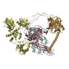
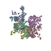
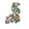
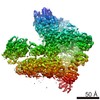
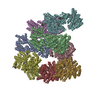
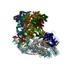
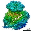
ジャーナル: Nature / 年: 2017 タイトル: Structure of the human MHC-I peptide-loading complex. 著者: Andreas Blees / Dovile Januliene / Tommy Hofmann / Nicole Koller / Carla Schmidt / Simon Trowitzsch / Arne Moeller / Robert Tampé / 要旨: The peptide-loading complex (PLC) is a transient, multisubunit membrane complex in the endoplasmic reticulum that is essential for establishing a hierarchical immune response. The PLC coordinates ...The peptide-loading complex (PLC) is a transient, multisubunit membrane complex in the endoplasmic reticulum that is essential for establishing a hierarchical immune response. The PLC coordinates peptide translocation into the endoplasmic reticulum with loading and editing of major histocompatibility complex class I (MHC-I) molecules. After final proofreading in the PLC, stable peptide-MHC-I complexes are released to the cell surface to evoke a T-cell response against infected or malignant cells. Sampling of different MHC-I allomorphs requires the precise coordination of seven different subunits in a single macromolecular assembly, including the transporter associated with antigen processing (TAP1 and TAP2, jointly referred to as TAP), the oxidoreductase ERp57, the MHC-I heterodimer, and the chaperones tapasin and calreticulin. The molecular organization of and mechanistic events that take place in the PLC are unknown owing to the heterogeneous composition and intrinsically dynamic nature of the complex. Here, we isolate human PLC from Burkitt's lymphoma cells using an engineered viral inhibitor as bait and determine the structure of native PLC by electron cryo-microscopy. Two endoplasmic reticulum-resident editing modules composed of tapasin, calreticulin, ERp57, and MHC-I are centred around TAP in a pseudo-symmetric orientation. A multivalent chaperone network within and across the editing modules establishes the proofreading function at two lateral binding platforms for MHC-I molecules. The lectin-like domain of calreticulin senses the MHC-I glycan, whereas the P domain reaches over the MHC-I peptide-binding pocket towards ERp57. This arrangement allows tapasin to facilitate peptide editing by clamping MHC-I. The translocation pathway of TAP opens out into a large endoplasmic reticulum lumenal cavity, confined by the membrane entry points of tapasin and MHC-I. Two lateral windows channel the antigenic peptides to MHC-I. Structures of PLC captured at distinct assembly states provide mechanistic insight into the recruitment and release of MHC-I. Our work defines the molecular symbiosis of an ABC transporter and an endoplasmic reticulum chaperone network in MHC-I assembly and provides insight into the onset of the adaptive immune response.
凍結剤: ETHANE / チャンバー内湿度: 100 % / チャンバー内温度: 277 K / 装置: FEI VITROBOT MARK IV
-
電子顕微鏡法
顕微鏡
FEI TITAN KRIOS
撮影
フィルム・検出器のモデル: GATAN K2 QUANTUM (4k x 4k) 平均電子線量: 55.0 e/Å2
電子線
加速電圧: 300 kV / 電子線源: FIELD EMISSION GUN
電子光学系
照射モード: FLOOD BEAM / 撮影モード: BRIGHT FIELD
実験機器
モデル: Titan Krios / 画像提供: FEI Company
+
画像解析
CTF補正
ソフトウェア - 名称: Gctf / 詳細: CTF correction was performed internally in Relion
初期モデル
モデルのタイプ: OTHER 詳細: Selected particles from the best 2D class averages were subjected to 3D-classification in Relion, using a low-pass filtered global average as a starting model. The best map was then used as ...詳細: Selected particles from the best 2D class averages were subjected to 3D-classification in Relion, using a low-pass filtered global average as a starting model. The best map was then used as an initial model for subsequent 3D classification.
 ムービー
ムービー コントローラー


 データを開く
 基本情報
基本情報 マップデータ
 試料
試料 Homo sapiens (ヒト)
 データ登録者
 引用
引用
 構造の表示
構造の表示 ムービービューア
 ダウンロードとリンク
 emd_3905.png
 http://ftp.pdbj.org/pub/emdb/structures/EMD-3905
 Z (Sec.)
 Y (Row.)
 X (Col.)


 試料の構成要素
 解析
解析 電子顕微鏡法
 FIELD EMISSION GUN