 Movie
Movie Controller


+ Open data

- Basic information
| Entry | Database: EMDB / ID: EMD-4396 | ||||||||||||||||||
|---|---|---|---|---|---|---|---|---|---|---|---|---|---|---|---|---|---|---|---|
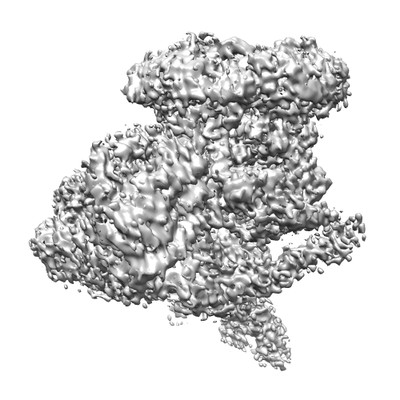
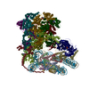
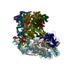
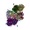
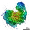
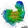
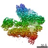
| Title | Chromatin remodeller-nucleosome complex at 4.5 A resolution. | ||||||||||||||||||
 Map data Map data | Chromatin remodeller-nucleosome complex at 4.5 A resolution. | ||||||||||||||||||
 Sample Sample |
| ||||||||||||||||||
 Keywords Keywords | Chromatin / Remodeller / ATPase / Histone / NUCLEAR PROTEIN | ||||||||||||||||||
| Function / homology |  Function and homology information Function and homology informationATP-dependent H2AZ histone chaperone activity / HDMs demethylate histones / sexual sporulation resulting in formation of a cellular spore / HATs acetylate histones / cupric reductase (NADH) activity / TTT Hsp90 cochaperone complex / Condensation of Prophase Chromosomes / RNA polymerase I upstream activating factor complex / global genome nucleotide-excision repair / R2TP complex ...ATP-dependent H2AZ histone chaperone activity / HDMs demethylate histones / sexual sporulation resulting in formation of a cellular spore / HATs acetylate histones / cupric reductase (NADH) activity / TTT Hsp90 cochaperone complex / Condensation of Prophase Chromosomes / RNA polymerase I upstream activating factor complex / global genome nucleotide-excision repair / R2TP complex / protein targeting to vacuole / Swr1 complex / SUMOylation of chromatin organization proteins / RMTs methylate histone arginines / Ino80 complex / DNA damage tolerance / rRNA transcription / box C/D snoRNP assembly / recombinational repair / kinetochore assembly / 5'-3' DNA helicase activity / NuA4 histone acetyltransferase complex / positive regulation of transcription by RNA polymerase I / intracellular copper ion homeostasis / mitotic metaphase chromosome alignment / nucleolar large rRNA transcription by RNA polymerase I / 3'-5' DNA helicase activity / CENP-A containing nucleosome / nucleosome binding / nuclear periphery / transcription initiation-coupled chromatin remodeling / aerobic respiration / kinetochore / Hydrolases; Acting on acid anhydrides; Acting on acid anhydrides to facilitate cellular and subcellular movement / rRNA processing / nucleosomal DNA binding / structural constituent of chromatin / nucleosome assembly / nucleosome / chromatin organization / heterochromatin formation / DNA helicase / molecular adaptor activity / DNA helicase activity / chromatin remodeling / protein stabilization / DNA repair / protein heterodimerization activity / regulation of transcription by RNA polymerase II / regulation of DNA-templated transcription / chromatin / negative regulation of transcription by RNA polymerase II / structural molecule activity / ATP hydrolysis activity / DNA binding / ATP binding / identical protein binding / nucleus / cytosol / cytoplasm Similarity search - Function | ||||||||||||||||||
| Biological species |   Saccharomyces cerevisiae (strain ATCC 204508 / S288c) (yeast) / synthetic construct (others) Saccharomyces cerevisiae (strain ATCC 204508 / S288c) (yeast) / synthetic construct (others) | ||||||||||||||||||
| Method | single particle reconstruction / cryo EM / Resolution: 3.6 Å | ||||||||||||||||||
 Authors Authors | Willhoft O / Chua EYD | ||||||||||||||||||
| Funding support |  United Kingdom, 5 items United Kingdom, 5 items
| ||||||||||||||||||
 Citation Citation | Journal: Science / Year: 2018 Title: Structure and dynamics of the yeast SWR1-nucleosome complex. Authors: Oliver Willhoft / Mohamed Ghoneim / Chia-Liang Lin / Eugene Y D Chua / Martin Wilkinson / Yuriy Chaban / Rafael Ayala / Elizabeth A McCormack / Lorraine Ocloo / David S Rueda / Dale B Wigley / Abstract: The yeast SWR1 complex exchanges histone H2A in nucleosomes with Htz1 (H2A.Z in humans). The cryo-electron microscopy structure of the SWR1 complex bound to a nucleosome at 3.6-angstrom resolution ...The yeast SWR1 complex exchanges histone H2A in nucleosomes with Htz1 (H2A.Z in humans). The cryo-electron microscopy structure of the SWR1 complex bound to a nucleosome at 3.6-angstrom resolution reveals details of the intricate interactions between components of the SWR1 complex and its nucleosome substrate. Interactions between the Swr1 motor domains and the DNA wrap at superhelical location 2 distort the DNA, causing a bulge with concomitant translocation of the DNA by one base pair, coupled to conformational changes of the histone core. Furthermore, partial unwrapping of the DNA from the histone core takes place upon binding of nucleosomes to SWR1 complex. The unwrapping, as monitored by single-molecule data, is stabilized and has its dynamics altered by adenosine triphosphate binding but does not require hydrolysis. | ||||||||||||||||||
| History |
|
- Structure visualization
| Movie |
Movie viewer |
|---|---|
| Structure viewer | EM map: SurfViewMolmilJmol/JSmol |
| Supplemental images |
- Downloads & links
-EMDB archive
| Map data | emd_4396.map.gz | 12.8 MB | EMDB map data format | |
|---|---|---|---|---|
| Header (meta data) | emd-4396-v30.xmlemd-4396.xml | 40.7 KB 40.7 KB | Display Display | EMDB header |
| Images |  emd_4396.png emd_4396.png | 67.7 KB | ||
| Filedesc metadata | emd-4396.cif.gz | 10.7 KB | ||
| Archive directory |  https://data.pdbj.org/pub/emdb/structures/EMD-4396ftp://data.pdbj.org/pub/emdb/structures/EMD-4396 https://data.pdbj.org/pub/emdb/structures/EMD-4396ftp://data.pdbj.org/pub/emdb/structures/EMD-4396 | HTTPS FTP |
-Related structure data
| Related structure data |  6genMC 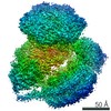 4395C  6gejC C: citing same article ( M: atomic model generated by this map |
|---|---|
| Similar structure data |

-Links
| EMDB pages | EMDB (EBI/PDBe) / EMDataResource |
|---|---|
| Related items in Molecule of the Month |





-Map
| File | Download / File: emd_4396.map.gz / Format: CCP4 / Size: 216 MB / Type: IMAGE STORED AS FLOATING POINT NUMBER (4 BYTES) | ||||||||||||||||||||||||||||||||||||||||||||||||||||||||||||||||||||
|---|---|---|---|---|---|---|---|---|---|---|---|---|---|---|---|---|---|---|---|---|---|---|---|---|---|---|---|---|---|---|---|---|---|---|---|---|---|---|---|---|---|---|---|---|---|---|---|---|---|---|---|---|---|---|---|---|---|---|---|---|---|---|---|---|---|---|---|---|---|
| Annotation | Chromatin remodeller-nucleosome complex at 4.5 A resolution. | ||||||||||||||||||||||||||||||||||||||||||||||||||||||||||||||||||||
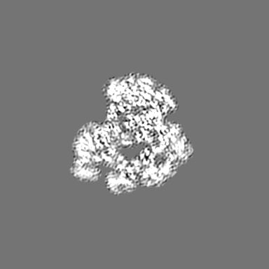
| Projections & slices | Image control
Images are generated by Spider. | ||||||||||||||||||||||||||||||||||||||||||||||||||||||||||||||||||||
| Voxel size | X=Y=Z: 1.11 Å | ||||||||||||||||||||||||||||||||||||||||||||||||||||||||||||||||||||
| Density |
| ||||||||||||||||||||||||||||||||||||||||||||||||||||||||||||||||||||
| Symmetry | Space group: 1 | ||||||||||||||||||||||||||||||||||||||||||||||||||||||||||||||||||||
| Details | EMDB XML:
CCP4 map header:
| ||||||||||||||||||||||||||||||||||||||||||||||||||||||||||||||||||||
 Z (Sec.)
 Y (Row.)
 X (Col.)




-Supplemental data
- Sample components
+Entire : SWR1-nucleosome complex
+Supramolecule #1: SWR1-nucleosome complex
+Supramolecule #2: Histones
+Supramolecule #3: DNA
+Supramolecule #4: remodeller
+Macromolecule #1: Vacuolar protein sorting-associated protein 72
 Trichoplusia ni (cabbage looper)
+Macromolecule #2: Histone H3

 Escherichia coli (E. coli)
+Macromolecule #3: Histone H4
+Macromolecule #4: Histone H2A.1
+Macromolecule #5: Histone H2B.1
+Macromolecule #8: Helicase SWR1
+Macromolecule #9: Actin-like protein ARP6
+Macromolecule #10: Vacuolar protein sorting-associated protein 71
+Macromolecule #11: RuvB-like protein 1
+Macromolecule #12: RuvB-like protein 2
+Macromolecule #6: DNA (173-MER)
+Macromolecule #7: DNA (173-MER)
+Macromolecule #13: ADENOSINE-5'-DIPHOSPHATE

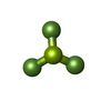
+Macromolecule #14: BERYLLIUM TRIFLUORIDE ION
+Macromolecule #15: MAGNESIUM ION
+Macromolecule #16: ZINC ION
-Experimental details
-Structure determination
| Method | cryo EM |
|---|---|
 Processing Processing | single particle reconstruction |
| Aggregation state | particle |
-Sample preparation
| Concentration | 0.03 mg/mL | ||||||||||||||||||
|---|---|---|---|---|---|---|---|---|---|---|---|---|---|---|---|---|---|---|---|
| Buffer | pH: 7 Component:
| ||||||||||||||||||
| Grid | Model: Quantifoil R1.2/1.3 / Material: GOLD / Mesh: 300 / Support film - Material: CARBON / Support film - topology: HOLEY / Pretreatment - Type: GLOW DISCHARGE / Pretreatment - Time: 5 sec. | ||||||||||||||||||
| Vitrification | Cryogen name: ETHANE / Chamber humidity: 100 % / Chamber temperature: 277.2 K / Instrument: FEI VITROBOT MARK IV |
- Electron microscopy
| Microscope | FEI TITAN KRIOS |
|---|---|
| Image recording | Film or detector model: FEI FALCON III (4k x 4k) / Detector mode: INTEGRATING / Number grids imaged: 1 / Number real images: 5517 / Average exposure time: 1.0 sec. / Average electron dose: 1.71794871794872 e/Å2 |
| Electron beam | Acceleration voltage: 300 kV / Electron source:  FIELD EMISSION GUN FIELD EMISSION GUN |
| Electron optics | Illumination mode: OTHER / Imaging mode: BRIGHT FIELD |
| Experimental equipment |  Model: Titan Krios / Image courtesy: FEI Company |
