 Movie
Movie Controller

[English] 日本語
 Yorodumi
- EMDB-37979: Human FL Metabotropic glutamate receptor 5, mGlu5-5M with quisqua... -
+ Open data

- Basic information
| Entry |  | ||||||||||||
|---|---|---|---|---|---|---|---|---|---|---|---|---|---|
| Title | Human FL Metabotropic glutamate receptor 5, mGlu5-5M with quisqualate, Rcc conformation | ||||||||||||
 Map data Map data | combined sharpened map, B=-70 | ||||||||||||
 Sample Sample |
| ||||||||||||
 Keywords Keywords | G-PROTEIN COUPLED RECEPTORS / SIGNAL TRANSDUCTION / METABOTROPIC GLUTAMATE RECEPTOR / Agonist / active state / MEMBRANE PROTEIN | ||||||||||||
| Function / homology |  Function and homology information Function and homology informationA2A adenosine receptor binding / sensory perception of hot stimulus / negative regulation of dendritic spine morphogenesis / : / G protein-coupled receptor activity involved in regulation of postsynaptic membrane potential / adenylate cyclase inhibiting G protein-coupled glutamate receptor activity / protein localization to nuclear inner membrane / positive regulation of cellular response to hypoxia / operant conditioning / phospholipase C-activating G protein-coupled glutamate receptor signaling pathway ...A2A adenosine receptor binding / sensory perception of hot stimulus / negative regulation of dendritic spine morphogenesis / : / G protein-coupled receptor activity involved in regulation of postsynaptic membrane potential / adenylate cyclase inhibiting G protein-coupled glutamate receptor activity / protein localization to nuclear inner membrane / positive regulation of cellular response to hypoxia / operant conditioning / phospholipase C-activating G protein-coupled glutamate receptor signaling pathway / positive regulation of long-term neuronal synaptic plasticity / positive regulation of sensory perception of pain / negative regulation of excitatory postsynaptic potential / desensitization of G protein-coupled receptor signaling pathway / positive regulation of dopamine secretion / G protein-coupled glutamate receptor signaling pathway / positive regulation of neural precursor cell proliferation / Class C/3 (Metabotropic glutamate/pheromone receptors) / glutamate receptor activity / nuclear inner membrane / Neurexins and neuroligins / astrocyte projection / response to corticosterone / response to morphine / temperature homeostasis / protein tyrosine kinase activator activity / conditioned place preference / regulation of synaptic transmission, glutamatergic / regulation of long-term synaptic depression / positive regulation of calcium-mediated signaling / protein tyrosine kinase binding / dendritic shaft / response to amphetamine / locomotory behavior / synapse organization / postsynaptic density membrane / Schaffer collateral - CA1 synapse / cognition / cellular response to amyloid-beta / G protein-coupled receptor activity / positive regulation of cytosolic calcium ion concentration / dendritic spine / G alpha (q) signalling events / chemical synaptic transmission / response to ethanol / learning or memory / positive regulation of MAPK cascade / response to antibiotic / neuronal cell body / regulation of DNA-templated transcription / dendrite / glutamatergic synapse / identical protein binding / plasma membrane / cytoplasm Similarity search - Function | ||||||||||||
| Biological species |  Homo sapiens (human) Homo sapiens (human) | ||||||||||||
| Method | single particle reconstruction / cryo EM / Resolution: 4.1 Å | ||||||||||||
 Authors Authors | Vinothkumar KR / Lebon G / Cannone G | ||||||||||||
| Funding support |  India, India,  France, 3 items France, 3 items
| ||||||||||||
 Citation Citation | Journal: Nat Commun / Year: 2025 Title: Conformational diversity in class C GPCR positive allosteric modulation. Authors: Giuseppe Cannone / Ludovic Berto / Fanny Malhaire / Gavin Ferguson / Aurelien Fouillen / Stéphanie Balor / Joan Font-Ingles / Amadeu Llebaria / Cyril Goudet / Abhay Kotecha / Vinothkumar K ...Authors: Giuseppe Cannone / Ludovic Berto / Fanny Malhaire / Gavin Ferguson / Aurelien Fouillen / Stéphanie Balor / Joan Font-Ingles / Amadeu Llebaria / Cyril Goudet / Abhay Kotecha / Vinothkumar K R / Guillaume Lebon /    Abstract: The metabotropic glutamate receptors (mGlus) are class C G protein-coupled receptors (GPCR) that form obligate dimers activated by the major excitatory neurotransmitter L-glutamate. The architecture ...The metabotropic glutamate receptors (mGlus) are class C G protein-coupled receptors (GPCR) that form obligate dimers activated by the major excitatory neurotransmitter L-glutamate. The architecture of mGlu receptor comprises an extracellular Venus-Fly Trap domain (VFT) connected to the transmembrane domain (7TM) through a Cysteine-Rich Domain (CRD). The binding of L-glutamate in the VFTs and subsequent conformational change results in the signal being transmitted to the 7TM inducing G protein binding and activation. The mGlu receptors signal transduction can be allosterically potentiated by positive allosteric modulators (PAMs) binding to the 7TMs, which are of therapeutic interest in various neurological disorders. Here, we report the cryoEM structures of metabotropic glutamate receptor 5 (mGlu) purified with three chemically and pharmacologically distinct PAMs. We find that the PAMs modulate the receptor equilibrium through their different binding modes, revealing how their interactions in the 7TMs impact the mGlu receptor conformational landscape and function. In addition, we identified a PAM-free but agonist-bound intermediate state that also reveals interactions mediated by intracellular loop 2. The activation of mGlu receptor is a multi-step process in which the binding of the PAMs in the 7TM modulates the equilibrium towards the active state. | ||||||||||||
| History |
|
- Structure visualization
| Supplemental images |
|---|

- Downloads & links
-EMDB archive
| Map data | emd_37979.map.gz | 254.7 MB | EMDB map data format | |
|---|---|---|---|---|
| Header (meta data) | emd-37979-v30.xmlemd-37979.xml | 21.5 KB 21.5 KB | Display Display | EMDB header |
| FSC (resolution estimation) | emd_37979_fsc.xml | 14.9 KB | Display | FSC data file |
| Images |  emd_37979.png emd_37979.png | 151.5 KB | ||
| Filedesc metadata | emd-37979.cif.gz | 7.5 KB | ||
| Others | emd_37979_half_map_1.map.gzemd_37979_half_map_2.map.gz | 254.6 MB 254.6 MB | ||
| Archive directory |  http://ftp.pdbj.org/pub/emdb/structures/EMD-37979ftp://ftp.pdbj.org/pub/emdb/structures/EMD-37979 http://ftp.pdbj.org/pub/emdb/structures/EMD-37979ftp://ftp.pdbj.org/pub/emdb/structures/EMD-37979 | HTTPS FTP |
-Related structure data
| Related structure data |  8x0hMC  8x0bC  8x0cC  8x0dC  8x0eC  8x0fC  8x0gC C: citing same article ( M: atomic model generated by this map |
|---|---|
| Similar structure data |


-Links
| EMDB pages | EMDB (EBI/PDBe) / EMDataResource |
|---|---|
| Related items in Molecule of the Month |


-Map
| File | Download / File: emd_37979.map.gz / Format: CCP4 / Size: 274.6 MB / Type: IMAGE STORED AS FLOATING POINT NUMBER (4 BYTES) | ||||||||||||||||||||||||||||||||||||
|---|---|---|---|---|---|---|---|---|---|---|---|---|---|---|---|---|---|---|---|---|---|---|---|---|---|---|---|---|---|---|---|---|---|---|---|---|---|
| Annotation | combined sharpened map, B=-70 | ||||||||||||||||||||||||||||||||||||
| Projections & slices | Image control
Images are generated by Spider. | ||||||||||||||||||||||||||||||||||||
| Voxel size | X=Y=Z: 0.84 Å | ||||||||||||||||||||||||||||||||||||
| Density |
| ||||||||||||||||||||||||||||||||||||
| Symmetry | Space group: 1 | ||||||||||||||||||||||||||||||||||||
| Details | EMDB XML:
|
 Z (Sec.)
 Y (Row.)
 X (Col.)


-Supplemental data
-Half map: one of the half maps
| File | emd_37979_half_map_1.map | ||||||||||||
|---|---|---|---|---|---|---|---|---|---|---|---|---|---|
| Annotation | one of the half maps | ||||||||||||
| Projections & Slices |
| ||||||||||||
| Density Histograms |


-Half map: one of the half maps
| File | emd_37979_half_map_2.map | ||||||||||||
|---|---|---|---|---|---|---|---|---|---|---|---|---|---|
| Annotation | one of the half maps | ||||||||||||
| Projections & Slices |
| ||||||||||||
| Density Histograms |


- Sample components
-Entire : mGlu Receptor bound to quisqualate, in Rcc conformation
| Entire | Name: mGlu Receptor bound to quisqualate, in Rcc conformation |
|---|---|
| Components |
|
-Supramolecule #1: mGlu Receptor bound to quisqualate, in Rcc conformation
| Supramolecule | Name: mGlu Receptor bound to quisqualate, in Rcc conformation type: complex / ID: 1 / Parent: 0 / Macromolecule list: #1 |
|---|---|
| Source (natural) | Organism: Homo sapiens (human) |
| Molecular weight | Theoretical: 200 KDa |
-Macromolecule #1: Metabotropic glutamate receptor 5
| Macromolecule | Name: Metabotropic glutamate receptor 5 / type: protein_or_peptide / ID: 1 Details: The construct has tags and protease cleavage site at its N-term (DYKDDDDKHHHHHHHHHHLEVLFQGP) and when compared to uniprot it starts at 21 and ends at 856. For CryoEM, the tag is cleaved and ...Details: The construct has tags and protease cleavage site at its N-term (DYKDDDDKHHHHHHHHHHLEVLFQGP) and when compared to uniprot it starts at 21 and ends at 856. For CryoEM, the tag is cleaved and the protein used for experiment starts at QSSE but residues starting RR have been modeled. There are 5 thermostabilising mutations (T742A, S753A, T777A, I799A, A813L). Residue N445 is mutated to remove glycosylation. An additional mutation H350L is engineered for a nanobody to bind. Each monomer has one sugar modeled and 10 disulfide bonds. Number of copies: 2 / Enantiomer: LEVO |
|---|---|
| Source (natural) | Organism: Homo sapiens (human) |
| Molecular weight | Theoretical: 93.691492 KDa |
| Recombinant expression | Organism: Homo sapiens (human) |
| Sequence | String: QSSERRVVAH MPGDIIIGAL FSVHHQPTVD KVHERKCGAV REQYGIQRVE AMLHTLERIN SDPTLLPNIT LGCEIRDSCW HSAVALEQS IEFIRDSLIS SEEEEGLVRC VDGSSSSFRS KKPIVGVIGP GSSSVAIQVQ NLLQLFNIPQ IAYSATSMDL S DKTLFKYF ...String: QSSERRVVAH MPGDIIIGAL FSVHHQPTVD KVHERKCGAV REQYGIQRVE AMLHTLERIN SDPTLLPNIT LGCEIRDSCW HSAVALEQS IEFIRDSLIS SEEEEGLVRC VDGSSSSFRS KKPIVGVIGP GSSSVAIQVQ NLLQLFNIPQ IAYSATSMDL S DKTLFKYF MRVVPSDAQQ ARAMVDIVKR YNWTYVSAVH TEGNYGESGM EAFKDMSAKE GICIAHSYKI YSNAGEQSFD KL LKKLTSH LPKARVVACF CEGMTVRGLL MAMRRLGLAG EFLLLGSDGW ADRYDVTDGY QREAVGGITI KLQSPDVKWF DDY YLKLRP ETNLRNPWFQ EFWQHRFQCR LEGFPQENSK YNKTCNSSLT LKTHHVQDSK MGFVINAIYS MAYGLHNMQM SLCP GYAGL CDAMKPIDGR KLLESLMKTA FTGVSGDTIL FDENGDSPGR YEIMNFKEMG KDYFDYINVG SWDNGELKMD DDEVW SKKS NIIRSVCSEP CEKGQIKVIR KGEVSCCWTC TPCKENEYVF DEYTCKACQL GSWPTDDLTG CDLIPVQYLR WGDPEP IAA VVFACLGLLA TLFVTVVFII YRDTPVVKSS SRELCYIILA GICLGYLCTF CLIAKPKQIY CYLQRIGIGL SPAMSYS AL VTKTNRIARI LAGSKKKICT KKPRFMSACA QLVIAFILIC IQLGIIVALF IMEPPDIMHD YPSIREVYLI CNTTNLGV V APLGYNGLLI LACTFYAFKT RNVPANFNEA KYIAFAMYTT CIIWLAFVPI YFGSNYKAIT MCFSVSLSAT VLLGCMFVP KVYIILAKPE RNVRSAFTTS TVVRMHVGDG KSSSAA UniProtKB: Metabotropic glutamate receptor 5 |
-Macromolecule #2: 2-acetamido-2-deoxy-beta-D-glucopyranose
| Macromolecule | Name: 2-acetamido-2-deoxy-beta-D-glucopyranose / type: ligand / ID: 2 / Number of copies: 2 / Formula: NAG |
|---|---|
| Molecular weight | Theoretical: 221.208 Da |
| Chemical component information |  ChemComp-NAG: |
-Macromolecule #3: (S)-2-AMINO-3-(3,5-DIOXO-[1,2,4]OXADIAZOLIDIN-2-YL)-PROPIONIC ACID
| Macromolecule | Name: (S)-2-AMINO-3-(3,5-DIOXO-[1,2,4]OXADIAZOLIDIN-2-YL)-PROPIONIC ACID type: ligand / ID: 3 / Number of copies: 2 / Formula: QUS |
|---|---|
| Molecular weight | Theoretical: 189.126 Da |
| Chemical component information | 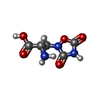 ChemComp-QUS: |
-Experimental details
-Structure determination
| Method | cryo EM |
|---|---|
 Processing Processing | single particle reconstruction |
| Aggregation state | particle |
-Sample preparation
| Concentration | 5 mg/mL | ||||||||||
|---|---|---|---|---|---|---|---|---|---|---|---|
| Buffer | pH: 7.4 Component:
| ||||||||||
| Grid | Model: Quantifoil R0.6/1 / Material: GOLD / Mesh: 300 / Support film - Material: CARBON / Support film - topology: HOLEY / Pretreatment - Type: GLOW DISCHARGE / Pretreatment - Time: 10 sec. / Pretreatment - Atmosphere: AIR | ||||||||||
| Vitrification | Cryogen name: ETHANE / Chamber humidity: 95 % / Chamber temperature: 277 K / Instrument: LEICA EM GP |
- Electron microscopy
| Microscope | FEI TITAN KRIOS |
|---|---|
| Details | Data was collected in EFTEM mode with Bioquantum K3 |
| Image recording | Film or detector model: GATAN K3 BIOQUANTUM (6k x 4k) / Digitization - Dimensions - Width: 5760 pixel / Digitization - Dimensions - Height: 4092 pixel / Number grids imaged: 1 / Number real images: 18710 / Average exposure time: 1.9 sec. / Average electron dose: 49.55 e/Å2 Details: Electron flux was 18.4 e/p/s and total number of frames were 50. |
| Electron beam | Acceleration voltage: 300 kV / Electron source:  FIELD EMISSION GUN FIELD EMISSION GUN |
| Electron optics | C2 aperture diameter: 50.0 µm / Calibrated magnification: 59523 / Illumination mode: FLOOD BEAM / Imaging mode: BRIGHT FIELD / Cs: 2.7 mm / Nominal defocus max: 2.2 µm / Nominal defocus min: 1.0 µm / Nominal magnification: 105000 |
| Sample stage | Specimen holder model: FEI TITAN KRIOS AUTOGRID HOLDER / Cooling holder cryogen: NITROGEN |
| Experimental equipment |  Model: Titan Krios / Image courtesy: FEI Company |
+Image processing

-Atomic model buiding 1
| Initial model |
| |||||||||
|---|---|---|---|---|---|---|---|---|---|---|
| Refinement | Space: REAL / Protocol: OTHER / Overall B value: 88.3 | |||||||||
| Output model | PDB-8x0h: |


