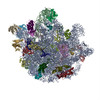
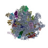
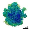
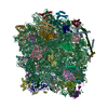
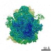
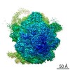
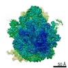
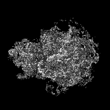
- EMDB-3748: Cryo-EM reconstruction of the small subunit of the Mycobacterium ... -
+
データを開く
IDまたはキーワード:
読み込み中...
-
基本情報
登録情報
データベース: EMDB / ID: EMD-3748
タイトル
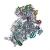
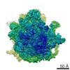
Cryo-EM reconstruction of the small subunit of the Mycobacterium smegmatis ribosome
マップデータ
試料
複合体: 30S small ribosomal subunit
RNA: x 3種
タンパク質・ペプチド: x 21種
リガンド: x 2種
キーワード
ribosome / translation
機能・相同性
機能・相同性情報
ribosomal small subunit assembly / ribosome biogenesis / ribosomal small subunit biogenesis / small ribosomal subunit / small ribosomal subunit rRNA binding / cytosolic small ribosomal subunit / tRNA binding / rRNA binding / structural constituent of ribosome / ribosome ...ribosomal small subunit assembly / ribosome biogenesis / ribosomal small subunit biogenesis / small ribosomal subunit / small ribosomal subunit rRNA binding / cytosolic small ribosomal subunit / tRNA binding / rRNA binding / structural constituent of ribosome / ribosome / translation / ribonucleoprotein complex / mRNA binding / RNA binding / zinc ion binding / metal ion binding / cytoplasm / cytosol 類似検索 - 分子機能
Mitochondrial mRNA-processing protein COX24, C-terminal / Mitochondrial mRNA-processing protein COX24, C-terminal / Mitochondrial domain of unknown function (DUF1713) / Ribosomal protein S14, type Z / Ribosomal protein L31 type A / Ribosomal protein S16, conserved site / Ribosomal protein S16 signature. / Ribosomal protein L31 signature. / Ribosomal protein L31 / Ribosomal protein L31 superfamily ...Mitochondrial mRNA-processing protein COX24, C-terminal / Mitochondrial mRNA-processing protein COX24, C-terminal / Mitochondrial domain of unknown function (DUF1713) / Ribosomal protein S14, type Z / Ribosomal protein L31 type A / Ribosomal protein S16, conserved site / Ribosomal protein S16 signature. / Ribosomal protein L31 signature. / Ribosomal protein L31 / Ribosomal protein L31 superfamily / Ribosomal protein L31 / Ribosomal protein S6, conserved site / Ribosomal protein S6 signature. / Ribosomal protein S3, bacterial-type / Ribosomal protein S13, bacterial-type / Ribosomal protein S19, bacterial-type / Ribosomal protein S7, bacterial/organellar-type / Ribosomal protein S11, bacterial-type / Ribosomal protein S20 / Ribosomal protein S20 superfamily / Ribosomal protein S20 / Ribosomal protein S4, bacterial-type / Ribosomal protein S5, bacterial-type / 30S ribosomal protein S17 / Ribosomal protein S6, plastid/chloroplast / Ribosomal protein S14/S29 / Ribosomal protein S2, bacteria/mitochondria/plastid / Ribosomal protein S18, conserved site / Ribosomal protein S18 signature. / Ribosomal protein S9, bacterial/plastid / Ribosomal protein S16 / Ribosomal protein S16 domain superfamily / Ribosomal protein S16 / L28p-like / Ribosomal protein S15, bacterial-type / Ribosomal protein S6 / Ribosomal protein S6 / Ribosomal protein S6 superfamily / Ribosomal protein S12, bacterial-type / Translation elongation factor EF1B/ribosomal protein S6 / Ribosomal protein S18 / Ribosomal protein S18 / Ribosomal protein S18 superfamily / K Homology domain / K homology RNA-binding domain / Ribosomal protein S3, conserved site / Ribosomal protein S3 signature. / Ribosomal protein S10, conserved site / Ribosomal protein S10 signature. / : / Ribosomal protein S14, conserved site / Ribosomal protein S14 signature. / Ribosomal protein S2 signature 1. / KH domain / Type-2 KH domain profile. / K Homology domain, type 2 / Ribosomal protein S3, C-terminal / Ribosomal protein S3, C-terminal domain / Ribosomal protein S3, C-terminal domain superfamily / Ribosomal protein S15/S19, conserved site / Ribosomal protein S19 signature. / Ribosomal protein S10 / Ribosomal protein S19/S15 / Ribosomal protein S19/S15, superfamily / Ribosomal protein S19 / Ribosomal protein S5, N-terminal, conserved site / Ribosomal protein S5 signature. / Ribosomal protein S7, conserved site / Ribosomal protein S7 signature. / Ribosomal protein S2, conserved site / : / K homology domain superfamily, prokaryotic type / Ribosomal protein S2 / Ribosomal protein S2, flavodoxin-like domain superfamily / Ribosomal protein S2 / Ribosomal protein S17, conserved site / Ribosomal protein S17 signature. / Ribosomal protein S5 / S5 double stranded RNA-binding domain profile. / Ribosomal protein S5, N-terminal / Ribosomal protein S13, conserved site / Ribosomal protein S13 signature. / Ribosomal protein S5, C-terminal / Ribosomal protein S5, N-terminal domain / Ribosomal protein S13 / 30s ribosomal protein S13, C-terminal / Ribosomal protein S13/S18 / Ribosomal protein S5, C-terminal domain / Ribosomal protein S13 family profile. / Ribosomal protein S4/S9 N-terminal domain / Ribosomal protein S8 signature. / Ribosomal protein S4, conserved site / Ribosomal protein S4 signature. / Ribosomal protein S4/S9 N-terminal domain / Ribosomal protein S4/S9, N-terminal / K homology domain-like, alpha/beta / Ribosomal protein S15 signature. / Ribosomal protein S14 / Ribosomal protein S14p/S29e / Ribosomal protein S4/S9 類似検索 - ドメイン・相同性
30S ribosomal protein S6 / Conserved domain protein / Small ribosomal subunit protein uS12 / Small ribosomal subunit protein uS7 / Small ribosomal subunit protein uS10 / Small ribosomal subunit protein uS19 / Small ribosomal subunit protein uS3 / Small ribosomal subunit protein uS17 / Small ribosomal subunit protein uS14B / Small ribosomal subunit protein uS8 ...30S ribosomal protein S6 / Conserved domain protein / Small ribosomal subunit protein uS12 / Small ribosomal subunit protein uS7 / Small ribosomal subunit protein uS10 / Small ribosomal subunit protein uS19 / Small ribosomal subunit protein uS3 / Small ribosomal subunit protein uS17 / Small ribosomal subunit protein uS14B / Small ribosomal subunit protein uS8 / Small ribosomal subunit protein uS5 / Small ribosomal subunit protein uS13 / Small ribosomal subunit protein uS11 / Small ribosomal subunit protein uS4 / Small ribosomal subunit protein uS9 / Small ribosomal subunit protein bS16 / Small ribosomal subunit protein uS2 / Small ribosomal subunit protein uS15 / Small ribosomal subunit protein bS20 / Large ribosomal subunit protein bL31 / Small ribosomal subunit protein bS18B 類似検索 - 構成要素
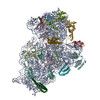
ジャーナル: Cell Rep / 年: 2017 タイトル: The Complete Structure of the Mycobacterium smegmatis 70S Ribosome. 著者: Jendrik Hentschel / Chloe Burnside / Ingrid Mignot / Marc Leibundgut / Daniel Boehringer / Nenad Ban / 要旨: The ribosome carries out the synthesis of proteins in every living cell. It consequently represents a frontline target in anti-microbial therapy. Tuberculosis ranks among the leading causes of death ...The ribosome carries out the synthesis of proteins in every living cell. It consequently represents a frontline target in anti-microbial therapy. Tuberculosis ranks among the leading causes of death worldwide, due in large part to the combination of difficult-to-treat latency and antibiotic resistance. Here, we present the 3.3-Å cryo-EM structure of the 70S ribosome of Mycobacterium smegmatis, a close relative to the human pathogen Mycobacterium tuberculosis. The structure reveals two additional ribosomal proteins and localizes them to the vicinity of drug-target sites in both the catalytic center and the decoding site of the ribosome. Furthermore, we visualized actinobacterium-specific rRNA and protein expansions that extensively remodel the ribosomal surface with implications for polysome organization. Our results provide a foundation for understanding the idiosyncrasies of mycobacterial translation and reveal atomic details of the structure that will facilitate the design of anti-tubercular therapeutics.
 ムービー
ムービー コントローラー


 データを開く
 基本情報
基本情報 マップデータ
 試料
試料 キーワード
キーワード 機能・相同性情報
 Mycobacterium smegmatis str. MC2 155 (バクテリア)
 データ登録者
 スイス, 2件
  引用
引用 構造の表示
構造の表示
 ダウンロードとリンク
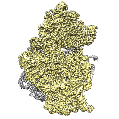 emd_3748.png
 http://ftp.pdbj.org/pub/emdb/structures/EMD-3748
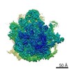


 Z (Sec.)
 Y (Row.)
 X (Col.)


 試料の構成要素
 解析
解析 電子顕微鏡法
 FIELD EMISSION GUN