 Movie
Movie Controller


+ Open data

- Basic information
| Entry | Database: EMDB / ID: EMD-31728 | |||||||||
|---|---|---|---|---|---|---|---|---|---|---|
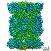
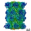
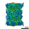
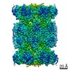
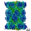
| Title | 20S proteasome incubated with monoUb-CyclinB1-NT (S0) | |||||||||
 Map data Map data | ||||||||||
 Sample Sample |
| |||||||||
| Biological species |  Homo sapiens (human) Homo sapiens (human) | |||||||||
| Method | single particle reconstruction / cryo EM / Resolution: 3.38 Å | |||||||||
 Authors Authors | Xu C / Cong Y | |||||||||
| Funding support |  Israel, 1 items Israel, 1 items
| |||||||||
 Citation Citation | Journal: Nat Commun / Year: 2021 Title: The 20S as a stand-alone proteasome in cells can degrade the ubiquitin tag. Authors: Indrajit Sahu / Sachitanand M Mali / Prasad Sulkshane / Cong Xu / Andrey Rozenberg / Roni Morag / Manisha Priyadarsini Sahoo / Sumeet K Singh / Zhanyu Ding / Yifan Wang / Sharleen Day / Yao ...Authors: Indrajit Sahu / Sachitanand M Mali / Prasad Sulkshane / Cong Xu / Andrey Rozenberg / Roni Morag / Manisha Priyadarsini Sahoo / Sumeet K Singh / Zhanyu Ding / Yifan Wang / Sharleen Day / Yao Cong / Oded Kleifeld / Ashraf Brik / Michael H Glickman /   Abstract: The proteasome, the primary protease for ubiquitin-dependent proteolysis in eukaryotes, is usually found as a mixture of 30S, 26S, and 20S complexes. These complexes have common catalytic sites, ...The proteasome, the primary protease for ubiquitin-dependent proteolysis in eukaryotes, is usually found as a mixture of 30S, 26S, and 20S complexes. These complexes have common catalytic sites, which makes it challenging to determine their distinctive roles in intracellular proteolysis. Here, we chemically synthesize a panel of homogenous ubiquitinated proteins, and use them to compare 20S and 26S proteasomes with respect to substrate selection and peptide-product generation. We show that 20S proteasomes can degrade the ubiquitin tag along with the conjugated substrate. Ubiquitin remnants on branched peptide products identified by LC-MS/MS, and flexibility in the 20S gate observed by cryo-EM, reflect the ability of the 20S proteasome to proteolyze an isopeptide-linked ubiquitin-conjugate. Peptidomics identifies proteasome-trapped ubiquitin-derived peptides and peptides of potential 20S substrates in Hi20S cells, hypoxic cells, and human failing-heart. Moreover, elevated levels of 20S proteasomes appear to contribute to cell survival under stress associated with damaged proteins. | |||||||||
| History |
|
- Structure visualization
| Movie |
 Movie viewer Movie viewer |
|---|---|
| Structure viewer | EM map: SurfViewMolmilJmol/JSmol |
| Supplemental images |
- Downloads & links
-EMDB archive
| Map data | emd_31728.map.gz | 28.5 MB | EMDB map data format | |
|---|---|---|---|---|
| Header (meta data) | emd-31728-v30.xmlemd-31728.xml | 9.7 KB 9.7 KB | Display Display | EMDB header |
| Images |  emd_31728.png emd_31728.png | 25.1 KB | ||
| Archive directory |  http://ftp.pdbj.org/pub/emdb/structures/EMD-31728ftp://ftp.pdbj.org/pub/emdb/structures/EMD-31728 http://ftp.pdbj.org/pub/emdb/structures/EMD-31728ftp://ftp.pdbj.org/pub/emdb/structures/EMD-31728 | HTTPS FTP |
-Validation report
| Summary document | emd_31728_validation.pdf.gz | 394.3 KB | Display | EMDB validaton report |
|---|---|---|---|---|
| Full document | emd_31728_full_validation.pdf.gz | 393.8 KB | Display | |
| Data in XML | emd_31728_validation.xml.gz | 5.7 KB | Display | |
| Data in CIF | emd_31728_validation.cif.gz | 6.5 KB | Display | |
| Arichive directory | https://ftp.pdbj.org/pub/emdb/validation_reports/EMD-31728ftp://ftp.pdbj.org/pub/emdb/validation_reports/EMD-31728 | HTTPS FTP |
-Related structure data


-Links
| EMDB pages | EMDB (EBI/PDBe) / EMDataResource |
|---|
-Map
| File | Download / File: emd_31728.map.gz / Format: CCP4 / Size: 30.5 MB / Type: IMAGE STORED AS FLOATING POINT NUMBER (4 BYTES) | ||||||||||||||||||||||||||||||||||||||||||||||||||||||||||||||||||||
|---|---|---|---|---|---|---|---|---|---|---|---|---|---|---|---|---|---|---|---|---|---|---|---|---|---|---|---|---|---|---|---|---|---|---|---|---|---|---|---|---|---|---|---|---|---|---|---|---|---|---|---|---|---|---|---|---|---|---|---|---|---|---|---|---|---|---|---|---|---|
| Projections & slices | Image control
Images are generated by Spider. | ||||||||||||||||||||||||||||||||||||||||||||||||||||||||||||||||||||
| Voxel size | X=Y=Z: 1.318 Å | ||||||||||||||||||||||||||||||||||||||||||||||||||||||||||||||||||||
| Density |
| ||||||||||||||||||||||||||||||||||||||||||||||||||||||||||||||||||||
| Symmetry | Space group: 1 | ||||||||||||||||||||||||||||||||||||||||||||||||||||||||||||||||||||
| Details | EMDB XML:
CCP4 map header:
| ||||||||||||||||||||||||||||||||||||||||||||||||||||||||||||||||||||
 Z (Sec.)
 Y (Row.)
 X (Col.)


-Supplemental data
- Sample components
-Entire : 20S proteasome incubated with monoUb CyclinB1-NT
| Entire | Name: 20S proteasome incubated with monoUb CyclinB1-NT |
|---|---|
| Components |
|
-Supramolecule #1: 20S proteasome incubated with monoUb CyclinB1-NT
| Supramolecule | Name: 20S proteasome incubated with monoUb CyclinB1-NT / type: complex / ID: 1 / Parent: 0 / Macromolecule list: #1-#14 |
|---|---|
| Source (natural) | Organism: Homo sapiens (human) |
-Experimental details
-Structure determination
| Method | cryo EM |
|---|---|
 Processing Processing | single particle reconstruction |
| Aggregation state | particle |
-Sample preparation
| Buffer | pH: 7.4 |
|---|---|
| Vitrification | Cryogen name: ETHANE |
- Electron microscopy
| Microscope | FEI TITAN KRIOS |
|---|---|
| Image recording | Film or detector model: GATAN K2 SUMMIT (4k x 4k) / Average electron dose: 38.0 e/Å2 |
| Electron beam | Acceleration voltage: 300 kV / Electron source:  FIELD EMISSION GUN FIELD EMISSION GUN |
| Electron optics | Illumination mode: FLOOD BEAM / Imaging mode: BRIGHT FIELD |
| Experimental equipment |  Model: Titan Krios / Image courtesy: FEI Company |
-Image processing
| Final reconstruction | Resolution.type: BY AUTHOR / Resolution: 3.38 Å / Resolution method: FSC 0.143 CUT-OFF / Number images used: 282834 |
|---|---|
| Initial angle assignment | Type: MAXIMUM LIKELIHOOD |
| Final angle assignment | Type: MAXIMUM LIKELIHOOD |
