 Movie
Movie Controller


+ Open data

- Basic information
| Entry | Database: PDB / ID: 7pg9 | |||||||||||||||||||||||||||
|---|---|---|---|---|---|---|---|---|---|---|---|---|---|---|---|---|---|---|---|---|---|---|---|---|---|---|---|---|
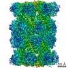
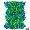
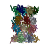
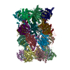
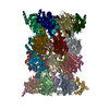
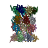
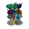
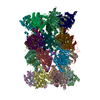
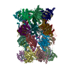
| Title | human 20S proteasome | |||||||||||||||||||||||||||
 Components Components |
| |||||||||||||||||||||||||||
 Keywords Keywords | HYDROLASE / 20S proteasome / human | |||||||||||||||||||||||||||
| Function / homology |  Function and homology information Function and homology informationpurine ribonucleoside triphosphate binding / Antigen processing: Ub, ATP-independent proteasomal degradation / sperm glycocalyx / Regulation of ornithine decarboxylase (ODC) / perinuclear theca / proteasome core complex / Proteasome assembly / Cross-presentation of soluble exogenous antigens (endosomes) / Somitogenesis / myofibril ...purine ribonucleoside triphosphate binding / Antigen processing: Ub, ATP-independent proteasomal degradation / sperm glycocalyx / Regulation of ornithine decarboxylase (ODC) / perinuclear theca / proteasome core complex / Proteasome assembly / Cross-presentation of soluble exogenous antigens (endosomes) / Somitogenesis / myofibril / proteasomal ubiquitin-independent protein catabolic process / sperm head-tail coupling apparatus / immune system process / proteasome endopeptidase complex / NF-kappaB binding / proteasome core complex, beta-subunit complex / threonine-type endopeptidase activity / proteasome core complex, alpha-subunit complex / proteasome assembly / : / ciliary tip / proteasome complex / sarcomere / Regulation of activated PAK-2p34 by proteasome mediated degradation / Autodegradation of Cdh1 by Cdh1:APC/C / centriole / APC/C:Cdc20 mediated degradation of Securin / negative regulation of inflammatory response to antigenic stimulus / sperm end piece / Asymmetric localization of PCP proteins / Ubiquitin-dependent degradation of Cyclin D / lipopolysaccharide binding / SCF-beta-TrCP mediated degradation of Emi1 / NIK-->noncanonical NF-kB signaling / AUF1 (hnRNP D0) binds and destabilizes mRNA / TNFR2 non-canonical NF-kB pathway / P-body / Assembly of the pre-replicative complex / Vpu mediated degradation of CD4 / Cdc20:Phospho-APC/C mediated degradation of Cyclin A / Dectin-1 mediated noncanonical NF-kB signaling / Degradation of DVL / Degradation of AXIN / Degradation of CRY and PER proteins / Hh mutants are degraded by ERAD / Activation of NF-kappaB in B cells / G2/M Checkpoints / Degradation of GLI1 by the proteasome / Hedgehog ligand biogenesis / Regulation of RUNX3 expression and activity / Autodegradation of the E3 ubiquitin ligase COP1 / Defective CFTR causes cystic fibrosis / GSK3B and BTRC:CUL1-mediated-degradation of NFE2L2 / Negative regulation of NOTCH4 signaling / APC/C:Cdh1 mediated degradation of Cdc20 and other APC/C:Cdh1 targeted proteins in late mitosis/early G1 / Hedgehog 'on' state / Vif-mediated degradation of APOBEC3G / FBXL7 down-regulates AURKA during mitotic entry and in early mitosis / Degradation of GLI2 by the proteasome / GLI3 is processed to GLI3R by the proteasome / Ubiquitin-Mediated Degradation of Phosphorylated Cdc25A / MAPK6/MAPK4 signaling / Degradation of CDH1 / Degradation of beta-catenin by the destruction complex / Oxygen-dependent proline hydroxylation of Hypoxia-inducible Factor Alpha / CDK-mediated phosphorylation and removal of Cdc6 / ABC-family protein mediated transport / CLEC7A (Dectin-1) signaling / SCF(Skp2)-mediated degradation of p27/p21 / response to virus / FCERI mediated NF-kB activation / nuclear matrix / Regulation of expression of SLITs and ROBOs / Regulation of PTEN stability and activity / Interleukin-1 signaling / Orc1 removal from chromatin / Regulation of RUNX2 expression and activity / Regulation of RAS by GAPs / The role of GTSE1 in G2/M progression after G2 checkpoint / Separation of Sister Chromatids / UCH proteinases / KEAP1-NFE2L2 pathway / peptidase activity / Downstream TCR signaling / Antigen processing: Ubiquitination & Proteasome degradation / sperm principal piece / RUNX1 regulates transcription of genes involved in differentiation of HSCs / ER-Phagosome pathway / Neddylation / regulation of inflammatory response / response to oxidative stress / sperm midpiece / secretory granule lumen / endopeptidase activity / ficolin-1-rich granule lumen / proteasome-mediated ubiquitin-dependent protein catabolic process / positive regulation of canonical NF-kappaB signal transduction / Ub-specific processing proteases / cilium / nuclear body Similarity search - Function | |||||||||||||||||||||||||||
| Biological species |  Homo sapiens (human) Homo sapiens (human) | |||||||||||||||||||||||||||
| Method | ELECTRON MICROSCOPY / single particle reconstruction / cryo EM / Resolution: 3.7 Å | |||||||||||||||||||||||||||
 Authors Authors | Xu, C. / Cong, Y. | |||||||||||||||||||||||||||
| Funding support |  Israel, 1items Israel, 1items
| |||||||||||||||||||||||||||
 Citation Citation | Journal: Nat Commun / Year: 2021 Title: The 20S as a stand-alone proteasome in cells can degrade the ubiquitin tag. Authors: Indrajit Sahu / Sachitanand M Mali / Prasad Sulkshane / Cong Xu / Andrey Rozenberg / Roni Morag / Manisha Priyadarsini Sahoo / Sumeet K Singh / Zhanyu Ding / Yifan Wang / Sharleen Day / Yao ...Authors: Indrajit Sahu / Sachitanand M Mali / Prasad Sulkshane / Cong Xu / Andrey Rozenberg / Roni Morag / Manisha Priyadarsini Sahoo / Sumeet K Singh / Zhanyu Ding / Yifan Wang / Sharleen Day / Yao Cong / Oded Kleifeld / Ashraf Brik / Michael H Glickman /   Abstract: The proteasome, the primary protease for ubiquitin-dependent proteolysis in eukaryotes, is usually found as a mixture of 30S, 26S, and 20S complexes. These complexes have common catalytic sites, ...The proteasome, the primary protease for ubiquitin-dependent proteolysis in eukaryotes, is usually found as a mixture of 30S, 26S, and 20S complexes. These complexes have common catalytic sites, which makes it challenging to determine their distinctive roles in intracellular proteolysis. Here, we chemically synthesize a panel of homogenous ubiquitinated proteins, and use them to compare 20S and 26S proteasomes with respect to substrate selection and peptide-product generation. We show that 20S proteasomes can degrade the ubiquitin tag along with the conjugated substrate. Ubiquitin remnants on branched peptide products identified by LC-MS/MS, and flexibility in the 20S gate observed by cryo-EM, reflect the ability of the 20S proteasome to proteolyze an isopeptide-linked ubiquitin-conjugate. Peptidomics identifies proteasome-trapped ubiquitin-derived peptides and peptides of potential 20S substrates in Hi20S cells, hypoxic cells, and human failing-heart. Moreover, elevated levels of 20S proteasomes appear to contribute to cell survival under stress associated with damaged proteins. | |||||||||||||||||||||||||||
| History |
|
- Structure visualization
| Movie |
Movie viewer |
|---|---|
| Structure viewer | Molecule: MolmilJmol/JSmol |
- Downloads & links
-Download
| PDBx/mmCIF format | 7pg9.cif.gz | 1013 KB | Display | PDBx/mmCIF format |
|---|---|---|---|---|
| PDB format | pdb7pg9.ent.gz | 815.5 KB | Display | PDB format |
| PDBx/mmJSON format | 7pg9.json.gz | Tree view | PDBx/mmJSON format | |
| Others |  Other downloads Other downloads |
-Validation report
| Arichive directory | https://data.pdbj.org/pub/pdb/validation_reports/pg/7pg9ftp://data.pdbj.org/pub/pdb/validation_reports/pg/7pg9 | HTTPS FTP |
|---|
-Related structure data
| Related structure data |  13389MC  7v5gC  7v5mC M: map data used to model this data C: citing same article ( |
|---|---|
| Similar structure data |


-Links
 PDBj
PDBj


- Assembly
| Deposited unit | 
|
|---|---|
| 1 |
|
-Components
-Proteasome subunit alpha type- ... , 7 types, 14 molecules AOBPCQDRESFTGU
| #1: Protein | Mass: 27432.459 Da / Num. of mol.: 2 / Source method: isolated from a natural source / Source: (natural) Homo sapiens (human) / References: UniProt: P60900#2: Protein | Mass: 25927.535 Da / Num. of mol.: 2 / Source method: isolated from a natural source / Source: (natural) Homo sapiens (human) / References: UniProt: P25787#3: Protein | Mass: 29525.842 Da / Num. of mol.: 2 / Source method: isolated from a natural source / Source: (natural) Homo sapiens (human) / References: UniProt: P25789#4: Protein | Mass: 27929.891 Da / Num. of mol.: 2 / Source method: isolated from a natural source / Source: (natural) Homo sapiens (human) / References: UniProt: O14818#5: Protein | Mass: 26435.977 Da / Num. of mol.: 2 / Source method: isolated from a natural source / Source: (natural) Homo sapiens (human) / References: UniProt: P28066#6: Protein | Mass: 29595.627 Da / Num. of mol.: 2 / Source method: isolated from a natural source / Source: (natural) Homo sapiens (human) / References: UniProt: P25786#7: Protein | Mass: 28469.252 Da / Num. of mol.: 2 / Source method: isolated from a natural source / Source: (natural) Homo sapiens (human) / References: UniProt: P25788 |
|---|
-Proteasome subunit beta type- ... , 7 types, 14 molecules HVIWJXKYLZMaNb
| #8: Protein | Mass: 21921.836 Da / Num. of mol.: 2 / Source method: isolated from a natural source / Source: (natural) Homo sapiens (human)References: UniProt: P28072, proteasome endopeptidase complex #9: Protein | Mass: 25321.980 Da / Num. of mol.: 2 / Source method: isolated from a natural source / Source: (natural) Homo sapiens (human)References: UniProt: Q99436, proteasome endopeptidase complex #10: Protein | Mass: 22972.896 Da / Num. of mol.: 2 / Source method: isolated from a natural source / Source: (natural) Homo sapiens (human)References: UniProt: P49720, proteasome endopeptidase complex #11: Protein | Mass: 22864.277 Da / Num. of mol.: 2 / Source method: isolated from a natural source / Source: (natural) Homo sapiens (human)References: UniProt: P49721, proteasome endopeptidase complex #12: Protein | Mass: 22484.369 Da / Num. of mol.: 2 / Source method: isolated from a natural source / Source: (natural) Homo sapiens (human)References: UniProt: P28074, proteasome endopeptidase complex #13: Protein | Mass: 23578.986 Da / Num. of mol.: 2 / Fragment: 20S CORE / Source method: isolated from a natural source / Source: (natural) Homo sapiens (human)References: UniProt: P20618, proteasome endopeptidase complex #14: Protein | Mass: 24414.740 Da / Num. of mol.: 2 / Source method: isolated from a natural source / Source: (natural) Homo sapiens (human)References: UniProt: P28070, proteasome endopeptidase complex |
|---|
-Details
| Has protein modification | N |
|---|
-Experimental details
-Experiment
| Experiment | Method: ELECTRON MICROSCOPY |
|---|---|
| EM experiment | Aggregation state: PARTICLE / 3D reconstruction method: single particle reconstruction |
- Sample preparation
| Component | Name: human 20S proteasome / Type: COMPLEX / Entity ID: all / Source: NATURAL |
|---|---|
| Source (natural) | Organism: Homo sapiens (human) |
| Buffer solution | pH: 7.4 |
| Specimen | Embedding applied: NO / Shadowing applied: NO / Staining applied: NO / Vitrification applied: YES |
| Vitrification | Cryogen name: ETHANE |
- Electron microscopy imaging
| Experimental equipment |  Model: Titan Krios / Image courtesy: FEI Company |
|---|---|
| Microscopy | Model: FEI TITAN KRIOS |
| Electron gun | Electron source:  FIELD EMISSION GUN / Accelerating voltage: 300 kV / Illumination mode: FLOOD BEAM FIELD EMISSION GUN / Accelerating voltage: 300 kV / Illumination mode: FLOOD BEAM |
| Electron lens | Mode: BRIGHT FIELD |
| Image recording | Electron dose: 38 e/Å2 / Film or detector model: GATAN K2 SUMMIT (4k x 4k) |
- Processing
| Software | Name: PHENIX / Version: 1.10.1_2155: / Classification: refinement | ||||||||||||||||||||||||
|---|---|---|---|---|---|---|---|---|---|---|---|---|---|---|---|---|---|---|---|---|---|---|---|---|---|
| EM software | Name: PHENIX / Category: model refinement | ||||||||||||||||||||||||
| CTF correction | Type: PHASE FLIPPING AND AMPLITUDE CORRECTION | ||||||||||||||||||||||||
| 3D reconstruction | Resolution: 3.7 Å / Resolution method: FSC 0.143 CUT-OFF / Num. of particles: 154436 / Symmetry type: POINT | ||||||||||||||||||||||||
| Refine LS restraints |
|
