 Movie
Movie Controller


+ Open data

- Basic information
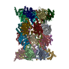
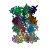
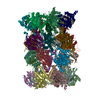
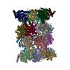
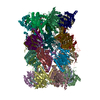
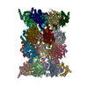
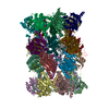
| Entry | Database: PDB / ID: 3wxr | ||||||
|---|---|---|---|---|---|---|---|
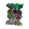
| Title | Yeast 20S proteasome with a mutation of alpha7 subunit | ||||||
 Components Components |
| ||||||
 Keywords Keywords | HYDROLASE / UPS / 20S proteasome / protease / 19S regulatory particle / MULTICATALYTIC PROTEASE | ||||||
| Function / homology |  Function and homology information Function and homology informationER-Phagosome pathway / Antigen processing: Ub, ATP-independent proteasomal degradation / proteasome core complex assembly / Proteasome assembly / Cross-presentation of soluble exogenous antigens (endosomes) / TNFR2 non-canonical NF-kB pathway / nuclear outer membrane-endoplasmic reticulum membrane network / Regulation of PTEN stability and activity / CDK-mediated phosphorylation and removal of Cdc6 / FBXL7 down-regulates AURKA during mitotic entry and in early mitosis ...ER-Phagosome pathway / Antigen processing: Ub, ATP-independent proteasomal degradation / proteasome core complex assembly / Proteasome assembly / Cross-presentation of soluble exogenous antigens (endosomes) / TNFR2 non-canonical NF-kB pathway / nuclear outer membrane-endoplasmic reticulum membrane network / Regulation of PTEN stability and activity / CDK-mediated phosphorylation and removal of Cdc6 / FBXL7 down-regulates AURKA during mitotic entry and in early mitosis / KEAP1-NFE2L2 pathway / Neddylation / Orc1 removal from chromatin / Ubiquitin-Mediated Degradation of Phosphorylated Cdc25A / MAPK6/MAPK4 signaling / Antigen processing: Ubiquitination & Proteasome degradation / proteasomal ubiquitin-independent protein catabolic process / Ub-specific processing proteases / proteasome storage granule / proteasome endopeptidase complex / proteasome core complex, beta-subunit complex / endopeptidase activator activity / threonine-type endopeptidase activity / proteasome core complex, alpha-subunit complex / proteasome assembly / Neutrophil degranulation / proteasome complex / peroxisome / endopeptidase activity / proteasome-mediated ubiquitin-dependent protein catabolic process / mRNA binding / endoplasmic reticulum membrane / mitochondrion / nucleus / cytosol Similarity search - Function | ||||||
| Biological species |  | ||||||
| Method |  X-RAY DIFFRACTION / SYNCHROTRON / MOLECULAR REPLACEMENT / Resolution: 3.15 Å X-RAY DIFFRACTION / SYNCHROTRON / MOLECULAR REPLACEMENT / Resolution: 3.15 Å | ||||||
 Authors Authors | Yashiroda, H. / Toda, Y. / Otsu, S. / Takagi, K. / Mizushima, T. / Murata, S. | ||||||
 Citation Citation | Journal: Mol.Cell.Biol. / Year: 2015 Title: N-terminal alpha 7 deletion of the proteasome 20S core particle substitutes for yeast PI31 function Authors: Yashiroda, H. / Toda, Y. / Otsu, S. / Takagi, K. / Mizushima, T. / Murata, S. | ||||||
| History |
|
- Structure visualization
| Structure viewer | Molecule: MolmilJmol/JSmol |
|---|
- Downloads & links
-Download
| PDBx/mmCIF format | 3wxr.cif.gz | 1.2 MB | Display | PDBx/mmCIF format |
|---|---|---|---|---|
| PDB format | pdb3wxr.ent.gz | 983.1 KB | Display | PDB format |
| PDBx/mmJSON format | 3wxr.json.gz | Tree view | PDBx/mmJSON format | |
| Others |  Other downloads Other downloads |
-Validation report
| Arichive directory | https://data.pdbj.org/pub/pdb/validation_reports/wx/3wxrftp://data.pdbj.org/pub/pdb/validation_reports/wx/3wxr | HTTPS FTP |
|---|
-Related structure data
| Related structure data | 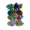 1rypS S: Starting model for refinement |
|---|---|
| Similar structure data |


-Links
 PDBj
PDBj


- Assembly
| Deposited unit | 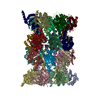
| ||||||||||||
|---|---|---|---|---|---|---|---|---|---|---|---|---|---|
| 1 |
| ||||||||||||
| Unit cell |
| ||||||||||||
| Noncrystallographic symmetry (NCS) | NCS oper:
|
-Components
-Proteasome subunit alpha type- ... , 6 types, 12 molecules AOBPCQDRESFT
| #1: Protein | Mass: 28033.830 Da / Num. of mol.: 2 / Source method: isolated from a natural source / Source: (natural) References: UniProt: P21243, proteasome endopeptidase complex #2: Protein | Mass: 27191.828 Da / Num. of mol.: 2 / Source method: isolated from a natural source / Source: (natural) References: UniProt: P23639, proteasome endopeptidase complex #3: Protein | Mass: 28748.230 Da / Num. of mol.: 2 / Source method: isolated from a natural source / Source: (natural) References: UniProt: P23638, proteasome endopeptidase complex #4: Protein | Mass: 28478.111 Da / Num. of mol.: 2 / Source method: isolated from a natural source / Source: (natural) References: UniProt: P40303, proteasome endopeptidase complex #5: Protein | Mass: 28649.086 Da / Num. of mol.: 2 / Source method: isolated from a natural source / Source: (natural) References: UniProt: P32379, proteasome endopeptidase complex #6: Protein | Mass: 25634.000 Da / Num. of mol.: 2 / Source method: isolated from a natural source / Source: (natural) References: UniProt: P40302, proteasome endopeptidase complex |
|---|
-Protein , 1 types, 2 molecules GU
| #7: Protein | Mass: 30465.934 Da / Num. of mol.: 2 / Fragment: UNP residues 13-288 / Source method: isolated from a natural source / Source: (natural) References: UniProt: P21242, proteasome endopeptidase complex |
|---|
-Proteasome subunit beta type- ... , 7 types, 14 molecules HVIWJXKYLZM1N2
| #8: Protein | Mass: 23573.604 Da / Num. of mol.: 2 / Source method: isolated from a natural source / Source: (natural) References: UniProt: P38624, proteasome endopeptidase complex #9: Protein | Mass: 28299.889 Da / Num. of mol.: 2 / Source method: isolated from a natural source / Source: (natural) References: UniProt: P25043, proteasome endopeptidase complex #10: Protein | Mass: 22627.842 Da / Num. of mol.: 2 / Source method: isolated from a natural source / Source: (natural) References: UniProt: P25451, proteasome endopeptidase complex #11: Protein | Mass: 26957.246 Da / Num. of mol.: 2 / Source method: isolated from a natural source / Source: (natural) References: UniProt: P22141, proteasome endopeptidase complex #12: Protein | Mass: 31670.539 Da / Num. of mol.: 2 / Source method: isolated from a natural source / Source: (natural) References: UniProt: P30656, proteasome endopeptidase complex #13: Protein | Mass: 26905.076 Da / Num. of mol.: 2 / Source method: isolated from a natural source / Source: (natural) References: UniProt: P23724, proteasome endopeptidase complex #14: Protein | Mass: 29471.289 Da / Num. of mol.: 2 / Source method: isolated from a natural source / Source: (natural) References: UniProt: P30657, proteasome endopeptidase complex |
|---|
-Experimental details
-Experiment
| Experiment | Method: X-RAY DIFFRACTION / Number of used crystals: 1 |
|---|
- Sample preparation
| Crystal | Density Matthews: 3.54 Å3/Da / Density % sol: 65.28 % |
|---|---|
| Crystal grow | Temperature: 293 K / Method: vapor diffusion, hanging drop / pH: 6.5 Details: 14% MPD, 0.1M MES, 50mM Magnesium acetate, pH 6.5, VAPOR DIFFUSION, HANGING DROP, temperature 293K |
-Data collection
| Diffraction | Mean temperature: 100 K |
|---|---|
| Diffraction source | Source: SYNCHROTRON / Site: SPring-8  / Beamline: BL44XU / Wavelength: 0.9 Å / Beamline: BL44XU / Wavelength: 0.9 Å |
| Detector | Type: RAYONIX MX300HE / Detector: CCD / Date: Jul 15, 2013 |
| Radiation | Protocol: SINGLE WAVELENGTH / Monochromatic (M) / Laue (L): M / Scattering type: x-ray |
| Radiation wavelength | Wavelength: 0.9 Å / Relative weight: 1 |
| Reflection | Resolution: 3.15→50 Å / Num. obs: 183783 / % possible obs: 87 % / Observed criterion σ(I): -3 / Redundancy: 3.4 % / Rmerge(I) obs: 0.135 / Net I/σ(I): 13.66 |
| Reflection shell | Resolution: 3.15→3.2 Å / Redundancy: 3.2 % / Rmerge(I) obs: 0.422 / Mean I/σ(I) obs: 3.461 / % possible all: 82.2 |
- Processing
| Software |
| ||||||||||||||||||||||||||||||||||||||||||||||||||||||||||||||||||||||||||||||||||||||||||||||||||||||||||||||||||||||||||||||||||||||||||||||||||||||||||||||||||||||||||||||||||||||
|---|---|---|---|---|---|---|---|---|---|---|---|---|---|---|---|---|---|---|---|---|---|---|---|---|---|---|---|---|---|---|---|---|---|---|---|---|---|---|---|---|---|---|---|---|---|---|---|---|---|---|---|---|---|---|---|---|---|---|---|---|---|---|---|---|---|---|---|---|---|---|---|---|---|---|---|---|---|---|---|---|---|---|---|---|---|---|---|---|---|---|---|---|---|---|---|---|---|---|---|---|---|---|---|---|---|---|---|---|---|---|---|---|---|---|---|---|---|---|---|---|---|---|---|---|---|---|---|---|---|---|---|---|---|---|---|---|---|---|---|---|---|---|---|---|---|---|---|---|---|---|---|---|---|---|---|---|---|---|---|---|---|---|---|---|---|---|---|---|---|---|---|---|---|---|---|---|---|---|---|---|---|---|---|
| Refinement | Method to determine structure: MOLECULAR REPLACEMENT Starting model: 1RYP Resolution: 3.15→48.2 Å / Cor.coef. Fo:Fc: 0.936 / Cor.coef. Fo:Fc free: 0.874 / SU B: 18.268 / SU ML: 0.307 / Cross valid method: THROUGHOUT / ESU R Free: 0.465 / Stereochemistry target values: MAXIMUM LIKELIHOOD / Details: HYDROGENS HAVE BEEN ADDED IN THE RIDING POSITIONS
| ||||||||||||||||||||||||||||||||||||||||||||||||||||||||||||||||||||||||||||||||||||||||||||||||||||||||||||||||||||||||||||||||||||||||||||||||||||||||||||||||||||||||||||||||||||||
| Solvent computation | Ion probe radii: 0.8 Å / Shrinkage radii: 0.8 Å / VDW probe radii: 1.2 Å / Solvent model: MASK | ||||||||||||||||||||||||||||||||||||||||||||||||||||||||||||||||||||||||||||||||||||||||||||||||||||||||||||||||||||||||||||||||||||||||||||||||||||||||||||||||||||||||||||||||||||||
| Displacement parameters | Biso mean: 51.662 Å2
| ||||||||||||||||||||||||||||||||||||||||||||||||||||||||||||||||||||||||||||||||||||||||||||||||||||||||||||||||||||||||||||||||||||||||||||||||||||||||||||||||||||||||||||||||||||||
| Refinement step | Cycle: LAST / Resolution: 3.15→48.2 Å
| ||||||||||||||||||||||||||||||||||||||||||||||||||||||||||||||||||||||||||||||||||||||||||||||||||||||||||||||||||||||||||||||||||||||||||||||||||||||||||||||||||||||||||||||||||||||
| Refine LS restraints |
|