 Movie
Movie Controller


+ Open data

- Basic information
| Entry | Database: PDB / ID: 4lqi | ||||||
|---|---|---|---|---|---|---|---|
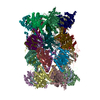
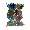
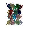
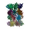
| Title | Yeast 20S Proteasome in complex with Vibralactone | ||||||
 Components Components |
| ||||||
 Keywords Keywords | HYDROLASE/HYDROLASE INHIBITOR / beta-lactone / inhibitor / natural product / target identification / Ntn-protease / cellular protein degradation / HYDROLASE-HYDROLASE INHIBITOR complex | ||||||
| Function / homology |  Function and homology information Function and homology informationER-Phagosome pathway / Antigen processing: Ub, ATP-independent proteasomal degradation / proteasome core complex assembly / Proteasome assembly / Cross-presentation of soluble exogenous antigens (endosomes) / TNFR2 non-canonical NF-kB pathway / nuclear outer membrane-endoplasmic reticulum membrane network / Regulation of PTEN stability and activity / CDK-mediated phosphorylation and removal of Cdc6 / FBXL7 down-regulates AURKA during mitotic entry and in early mitosis ...ER-Phagosome pathway / Antigen processing: Ub, ATP-independent proteasomal degradation / proteasome core complex assembly / Proteasome assembly / Cross-presentation of soluble exogenous antigens (endosomes) / TNFR2 non-canonical NF-kB pathway / nuclear outer membrane-endoplasmic reticulum membrane network / Regulation of PTEN stability and activity / CDK-mediated phosphorylation and removal of Cdc6 / FBXL7 down-regulates AURKA during mitotic entry and in early mitosis / KEAP1-NFE2L2 pathway / Neddylation / Orc1 removal from chromatin / Ubiquitin-Mediated Degradation of Phosphorylated Cdc25A / MAPK6/MAPK4 signaling / Antigen processing: Ubiquitination & Proteasome degradation / proteasomal ubiquitin-independent protein catabolic process / Ub-specific processing proteases / proteasome storage granule / proteasome endopeptidase complex / proteasome core complex, beta-subunit complex / endopeptidase activator activity / threonine-type endopeptidase activity / proteasome core complex, alpha-subunit complex / proteasome assembly / Neutrophil degranulation / proteasome complex / peroxisome / endopeptidase activity / proteasome-mediated ubiquitin-dependent protein catabolic process / mRNA binding / endoplasmic reticulum membrane / mitochondrion / nucleus / cytosol Similarity search - Function | ||||||
| Biological species |  | ||||||
| Method |  X-RAY DIFFRACTION / SYNCHROTRON / MOLECULAR REPLACEMENT / Resolution: 2.7 Å X-RAY DIFFRACTION / SYNCHROTRON / MOLECULAR REPLACEMENT / Resolution: 2.7 Å | ||||||
 Authors Authors | List, A. / Zeiler, E. / Gallastegui, N. / Rusch, M. / Hedberg, C. / Sieber, S.A. / Groll, M. | ||||||
 Citation Citation | Journal: Angew.Chem.Int.Ed.Engl. / Year: 2014 Title: Omuralide and Vibralactone: Differences in the Proteasome-beta-Lactone-gamma-Lactam Binding Scaffold Alter Target Preferences. Authors: List, A. / Zeiler, E. / Gallastegui, N. / Rusch, M. / Hedberg, C. / Sieber, S.A. / Groll, M. | ||||||
| History |
|
- Structure visualization
| Structure viewer | Molecule: MolmilJmol/JSmol |
|---|
- Downloads & links
-Download
| PDBx/mmCIF format | 4lqi.cif.gz | 1.2 MB | Display | PDBx/mmCIF format |
|---|---|---|---|---|
| PDB format | pdb4lqi.ent.gz | 1012.7 KB | Display | PDB format |
| PDBx/mmJSON format | 4lqi.json.gz | Tree view | PDBx/mmJSON format | |
| Others |  Other downloads Other downloads |
-Validation report
| Arichive directory | https://data.pdbj.org/pub/pdb/validation_reports/lq/4lqiftp://data.pdbj.org/pub/pdb/validation_reports/lq/4lqi | HTTPS FTP |
|---|
-Related structure data
| Related structure data |  1rypS S: Starting model for refinement |
|---|---|
| Similar structure data |


-Links
 PDBj
PDBj


- Assembly
| Deposited unit | 
| ||||||||
|---|---|---|---|---|---|---|---|---|---|
| 1 |
| ||||||||
| Unit cell |
|
-Components
-Proteasome subunit alpha type- ... , 7 types, 14 molecules AOBPCQDRESFTGU
| #1: Protein | Mass: 27191.828 Da / Num. of mol.: 2 / Source method: isolated from a natural source / Source: (natural) References: UniProt: P23639, proteasome endopeptidase complex #2: Protein | Mass: 27050.416 Da / Num. of mol.: 2 / Source method: isolated from a natural source / Source: (natural) References: UniProt: P23638, proteasome endopeptidase complex #3: Protein | Mass: 26903.330 Da / Num. of mol.: 2 / Source method: isolated from a natural source / Source: (natural) References: UniProt: P40303, proteasome endopeptidase complex #4: Protein | Mass: 26544.789 Da / Num. of mol.: 2 / Source method: isolated from a natural source / Source: (natural) References: UniProt: P32379, proteasome endopeptidase complex #5: Protein | Mass: 25502.805 Da / Num. of mol.: 2 / Source method: isolated from a natural source / Source: (natural) References: UniProt: P40302, proteasome endopeptidase complex #6: Protein | Mass: 26892.482 Da / Num. of mol.: 2 / Source method: isolated from a natural source / Source: (natural) References: UniProt: P21242, proteasome endopeptidase complex #7: Protein | Mass: 27316.037 Da / Num. of mol.: 2 / Source method: isolated from a natural source / Source: (natural) References: UniProt: P21243, proteasome endopeptidase complex |
|---|
-Proteasome subunit beta type- ... , 7 types, 14 molecules HVIWJXKYLZMaNb
| #8: Protein | Mass: 23987.254 Da / Num. of mol.: 2 / Source method: isolated from a natural source / Source: (natural) References: UniProt: P25043, proteasome endopeptidase complex #9: Protein | Mass: 22496.645 Da / Num. of mol.: 2 / Source method: isolated from a natural source / Source: (natural) References: UniProt: P25451, proteasome endopeptidase complex #10: Protein | Mass: 22545.676 Da / Num. of mol.: 2 / Source method: isolated from a natural source / Source: (natural) References: UniProt: P22141, proteasome endopeptidase complex #11: Protein | Mass: 23325.248 Da / Num. of mol.: 2 / Source method: isolated from a natural source / Source: (natural) References: UniProt: P30656, proteasome endopeptidase complex #12: Protein | Mass: 24883.928 Da / Num. of mol.: 2 / Source method: isolated from a natural source / Source: (natural) References: UniProt: P23724, proteasome endopeptidase complex #13: Protein | Mass: 25945.496 Da / Num. of mol.: 2 / Source method: isolated from a natural source / Source: (natural) References: UniProt: P30657, proteasome endopeptidase complex #14: Protein | Mass: 21517.186 Da / Num. of mol.: 2 / Source method: isolated from a natural source / Source: (natural) References: UniProt: P38624, proteasome endopeptidase complex |
|---|
-Non-polymers , 2 types, 1338 molecules 

| #15: Chemical | ChemComp-1Y9 /  Mass: 210.270 Da / Num. of mol.: 4 / Source method: obtained synthetically / Formula: C12H18O3 Mass: 210.270 Da / Num. of mol.: 4 / Source method: obtained synthetically / Formula: C12H18O3#16: Water | ChemComp-HOH / | Mass: 18.015 Da / Num. of mol.: 1334 / Source method: isolated from a natural source / Formula: H2O |
|---|
-Details
| Has protein modification | Y |
|---|
-Experimental details
-Experiment
| Experiment | Method: X-RAY DIFFRACTION / Number of used crystals: 1 |
|---|
- Sample preparation
| Crystal | Density Matthews: 3.85 Å3/Da / Density % sol: 68.07 % |
|---|---|
| Crystal grow | Temperature: 293 K / Method: vapor diffusion, hanging drop / pH: 7 Details: 20 mM magnesium acetate, 100 mM MES, 12% MPD, pH 7.0, VAPOR DIFFUSION, HANGING DROP, temperature 293K |
-Data collection
| Diffraction | Mean temperature: 100 K |
|---|---|
| Diffraction source | Source: SYNCHROTRON / Site: SLS  / Beamline: X06SA / Wavelength: 1 Å / Beamline: X06SA / Wavelength: 1 Å |
| Detector | Type: PSI PILATUS 6M / Detector: PIXEL / Date: Aug 16, 2011 |
| Radiation | Monochromator: LN2 cooled fixed-exit Si(111) / Protocol: SINGLE WAVELENGTH / Monochromatic (M) / Laue (L): M / Scattering type: x-ray |
| Radiation wavelength | Wavelength: 1 Å / Relative weight: 1 |
| Reflection | Resolution: 2.7→40 Å / Num. all: 290996 / Num. obs: 289175 / % possible obs: 99.4 % / Observed criterion σ(F): 2 / Observed criterion σ(I): 2 / Redundancy: 3.4 % / Biso Wilson estimate: 46.3 Å2 / Rmerge(I) obs: 0.113 / Net I/σ(I): 7.1 |
| Reflection shell | Resolution: 2.7→2.8 Å / Rmerge(I) obs: 0.523 / Mean I/σ(I) obs: 1.8 / % possible all: 99.7 |
- Processing
| Software |
| |||||||||||||||||||||||||
|---|---|---|---|---|---|---|---|---|---|---|---|---|---|---|---|---|---|---|---|---|---|---|---|---|---|---|
| Refinement | Method to determine structure: MOLECULAR REPLACEMENT Starting model: PDB ENTRY 1RYP Resolution: 2.7→14.99 Å / Rfactor Rfree error: 0.002 / Data cutoff high absF: 6031462.9 / Data cutoff low absF: 0 / Isotropic thermal model: RESTRAINED / Cross valid method: THROUGHOUT / σ(F): 0 / Details: BULK SOLVENT MODEL USED
| |||||||||||||||||||||||||
| Solvent computation | Solvent model: FLAT MODEL / Bsol: 43.4917 Å2 / ksol: 0.35 e/Å3 | |||||||||||||||||||||||||
| Displacement parameters | Biso mean: 62.5 Å2
| |||||||||||||||||||||||||
| Refine analyze |
| |||||||||||||||||||||||||
| Refinement step | Cycle: LAST / Resolution: 2.7→14.99 Å
| |||||||||||||||||||||||||
| Refine LS restraints |
| |||||||||||||||||||||||||
| LS refinement shell | Resolution: 2.7→2.87 Å / Rfactor Rfree error: 0.007 / Total num. of bins used: 6
| |||||||||||||||||||||||||
| Xplor file |
|
