 Movie
Movie Controller

[English] 日本語
 Yorodumi
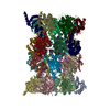
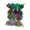
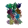
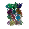
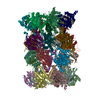
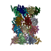
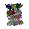
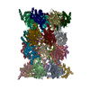
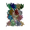
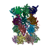
- PDB-1iru: Crystal Structure of the mammalian 20S proteasome at 2.75 A resolution -
+ Open data

- Basic information
| Entry | Database: PDB / ID: 1iru | ||||||
|---|---|---|---|---|---|---|---|
| Title | Crystal Structure of the mammalian 20S proteasome at 2.75 A resolution | ||||||
 Components Components | (20S proteasome) x 14 | ||||||
 Keywords Keywords | HYDROLASE / 20S proteasome / cell cycle / Immune response / proteolysis / ubiquitin | ||||||
| Function / homology |  Function and homology information Function and homology informationAntigen processing: Ub, ATP-independent proteasomal degradation / Regulation of ornithine decarboxylase (ODC) / Proteasome assembly / Cross-presentation of soluble exogenous antigens (endosomes) / ER-Phagosome pathway / Autodegradation of Cdh1 by Cdh1:APC/C / APC/C:Cdc20 mediated degradation of Securin / APC/C:Cdh1 mediated degradation of Cdc20 and other APC/C:Cdh1 targeted proteins in late mitosis/early G1 / Cdc20:Phospho-APC/C mediated degradation of Cyclin A / Autodegradation of the E3 ubiquitin ligase COP1 ...Antigen processing: Ub, ATP-independent proteasomal degradation / Regulation of ornithine decarboxylase (ODC) / Proteasome assembly / Cross-presentation of soluble exogenous antigens (endosomes) / ER-Phagosome pathway / Autodegradation of Cdh1 by Cdh1:APC/C / APC/C:Cdc20 mediated degradation of Securin / APC/C:Cdh1 mediated degradation of Cdc20 and other APC/C:Cdh1 targeted proteins in late mitosis/early G1 / Cdc20:Phospho-APC/C mediated degradation of Cyclin A / Autodegradation of the E3 ubiquitin ligase COP1 / Asymmetric localization of PCP proteins / Degradation of AXIN / Degradation of DVL / Hedgehog ligand biogenesis / Hedgehog 'on' state / TNFR2 non-canonical NF-kB pathway / Assembly of the pre-replicative complex / CDK-mediated phosphorylation and removal of Cdc6 / G2/M Checkpoints / Ubiquitin-Mediated Degradation of Phosphorylated Cdc25A / Ubiquitin-dependent degradation of Cyclin D / RUNX1 regulates transcription of genes involved in differentiation of HSCs / Regulation of RUNX3 expression and activity / Regulation of PTEN stability and activity / KEAP1-NFE2L2 pathway / Degradation of CDH1 / Degradation of CRY and PER proteins / Activation of NF-kappaB in B cells / Oxygen-dependent proline hydroxylation of Hypoxia-inducible Factor Alpha / SCF(Skp2)-mediated degradation of p27/p21 / FCERI mediated NF-kB activation / Dectin-1 mediated noncanonical NF-kB signaling / CLEC7A (Dectin-1) signaling / Degradation of GLI1 by the proteasome / NIK-->noncanonical NF-kB signaling / Orc1 removal from chromatin / FBXL7 down-regulates AURKA during mitotic entry and in early mitosis / Regulation of RUNX2 expression and activity / Interleukin-1 signaling / GSK3B and BTRC:CUL1-mediated-degradation of NFE2L2 / Degradation of beta-catenin by the destruction complex / Neddylation / Antigen processing: Ubiquitination & Proteasome degradation / UCH proteinases / Downstream TCR signaling / The role of GTSE1 in G2/M progression after G2 checkpoint / AUF1 (hnRNP D0) binds and destabilizes mRNA / Ub-specific processing proteases / ABC-family protein mediated transport / Separation of Sister Chromatids / MAPK6/MAPK4 signaling / GLI3 is processed to GLI3R by the proteasome / Neutrophil degranulation / proteasome core complex / immune system process / proteasome endopeptidase complex / proteasome core complex, beta-subunit complex / threonine-type endopeptidase activity / proteasome core complex, alpha-subunit complex / : / P-body / response to oxidative stress / endopeptidase activity / proteasome-mediated ubiquitin-dependent protein catabolic process / ciliary basal body / centrosome / mitochondrion / nucleoplasm / nucleus / cytoplasm / cytosol Similarity search - Function | ||||||
| Biological species |  | ||||||
| Method |  X-RAY DIFFRACTION / SYNCHROTRON / MOLECULAR REPLACEMENT / Resolution: 2.75 Å X-RAY DIFFRACTION / SYNCHROTRON / MOLECULAR REPLACEMENT / Resolution: 2.75 Å | ||||||
 Authors Authors | Unno, M. / Mizushima, T. / Morimoto, Y. / Tomisugi, Y. / Tanaka, K. / Yasuoka, N. / Tsukihara, T. | ||||||
 Citation Citation | Journal: Structure / Year: 2002 Title: The structure of the mammalian 20S proteasome at 2.75 A resolution. Authors: Unno, M. / Mizushima, T. / Morimoto, Y. / Tomisugi, Y. / Tanaka, K. / Yasuoka, N. / Tsukihara, T. | ||||||
| History |
|
- Structure visualization
| Structure viewer | Molecule: MolmilJmol/JSmol |
|---|
- Downloads & links
-Download
| PDBx/mmCIF format | 1iru.cif.gz | 1.2 MB | Display | PDBx/mmCIF format |
|---|---|---|---|---|
| PDB format | pdb1iru.ent.gz | 953 KB | Display | PDB format |
| PDBx/mmJSON format | 1iru.json.gz | Tree view | PDBx/mmJSON format | |
| Others |  Other downloads Other downloads |
-Validation report
| Arichive directory | https://data.pdbj.org/pub/pdb/validation_reports/ir/1iruftp://data.pdbj.org/pub/pdb/validation_reports/ir/1iru | HTTPS FTP |
|---|
-Related structure data
| Similar structure data |
|---|


-Links
 PDBj
PDBj


- Assembly
| Deposited unit | 
| ||||||||
|---|---|---|---|---|---|---|---|---|---|
| 1 |
| ||||||||
| Unit cell |
| ||||||||
| Details | 28mer |
-Components
-Protein , 14 types, 28 molecules AOBPCQDRESFTGUHVIWJXKYLZM1N2
| #1: Protein | Mass: 27432.459 Da / Num. of mol.: 2 / Source method: isolated from a natural source / Source: (natural) #2: Protein | Mass: 25796.338 Da / Num. of mol.: 2 / Source method: isolated from a natural source / Source: (natural) #3: Protein | Mass: 29525.842 Da / Num. of mol.: 2 / Source method: isolated from a natural source / Source: (natural) #4: Protein | Mass: 27929.891 Da / Num. of mol.: 2 / Source method: isolated from a natural source / Source: (natural) #5: Protein | Mass: 26494.016 Da / Num. of mol.: 2 / Source method: isolated from a natural source / Source: (natural) #6: Protein | Mass: 29595.627 Da / Num. of mol.: 2 / Source method: isolated from a natural source / Source: (natural) #7: Protein | Mass: 28338.057 Da / Num. of mol.: 2 / Source method: isolated from a natural source / Source: (natural) #8: Protein | Mass: 21921.836 Da / Num. of mol.: 2 / Source method: isolated from a natural source / Source: (natural) #9: Protein | Mass: 25351.047 Da / Num. of mol.: 2 / Source method: isolated from a natural source / Source: (natural) #10: Protein | Mass: 22954.859 Da / Num. of mol.: 2 / Source method: isolated from a natural source / Source: (natural) #11: Protein | Mass: 22864.277 Da / Num. of mol.: 2 / Source method: isolated from a natural source / Source: (natural) #12: Protein | Mass: 22484.369 Da / Num. of mol.: 2 / Source method: isolated from a natural source / Source: (natural) #13: Protein | Mass: 23578.986 Da / Num. of mol.: 2 / Source method: isolated from a natural source / Source: (natural) #14: Protein | Mass: 24402.686 Da / Num. of mol.: 2 / Source method: isolated from a natural source / Source: (natural) |
|---|
-Non-polymers , 2 types, 195 molecules 

| #15: Chemical | ChemComp-MG /  Mass: 24.305 Da / Num. of mol.: 30 / Source method: obtained synthetically / Formula: Mg Mass: 24.305 Da / Num. of mol.: 30 / Source method: obtained synthetically / Formula: Mg#16: Water | ChemComp-HOH / | Mass: 18.015 Da / Num. of mol.: 165 / Source method: isolated from a natural source / Formula: H2O |
|---|
-Details
| Has protein modification | Y |
|---|
-Experimental details
-Experiment
| Experiment | Method: X-RAY DIFFRACTION / Number of used crystals: 1 |
|---|
- Sample preparation
| Crystal | Density Matthews: 2.64 Å3/Da / Density % sol: 53.33 % | ||||||||||||||||||||||||
|---|---|---|---|---|---|---|---|---|---|---|---|---|---|---|---|---|---|---|---|---|---|---|---|---|---|
| Crystal grow | Temperature: 293 K / Method: vapor diffusion, hanging drop / pH: 6.5 Details: MPD, Mg acetate, Na cacodylate, pH 6.5, VAPOR DIFFUSION, HANGING DROP, temperature 293K | ||||||||||||||||||||||||
| Crystal grow | *PLUS Temperature: 25 ℃ / Details: Tomisugi, Y., (2000) J. Biochem., 127, 941. | ||||||||||||||||||||||||
| Components of the solutions | *PLUS
|
-Data collection
| Diffraction | Mean temperature: 100 K |
|---|---|
| Diffraction source | Source: SYNCHROTRON / Site: SPring-8  / Beamline: BL41XU / Wavelength: 1 Å / Beamline: BL41XU / Wavelength: 1 Å |
| Detector | Type: MARRESEARCH / Detector: CCD |
| Radiation | Monochromator: the rotated-inclined double-crystal monochrometer Protocol: SINGLE WAVELENGTH / Monochromatic (M) / Laue (L): M / Scattering type: x-ray |
| Radiation wavelength | Wavelength: 1 Å / Relative weight: 1 |
| Reflection | Resolution: 2.75→72.55 Å / Num. all: 189429 / Num. obs: 189429 / % possible obs: 96.3 % / Observed criterion σ(F): 0 / Observed criterion σ(I): 0 |
| Reflection shell | Resolution: 2.75→2.82 Å / % possible all: 70 |
| Reflection | *PLUS Lowest resolution: 100 Å / Num. measured all: 733250 / Rmerge(I) obs: 0.095 |
| Reflection shell | *PLUS % possible obs: 70 % / Rmerge(I) obs: 0.406 / Mean I/σ(I) obs: 1.8 |
- Processing
| Software |
| |||||||||||||||||||||||||
|---|---|---|---|---|---|---|---|---|---|---|---|---|---|---|---|---|---|---|---|---|---|---|---|---|---|---|
| Refinement | Method to determine structure: MOLECULAR REPLACEMENT / Resolution: 2.75→65 Å / σ(F): 0 / Stereochemistry target values: Engh & Huber
| |||||||||||||||||||||||||
| Refinement step | Cycle: LAST / Resolution: 2.75→65 Å
| |||||||||||||||||||||||||
| Refine LS restraints |
| |||||||||||||||||||||||||
| Refinement | *PLUS % reflection Rfree: 5 % / Rfactor obs: 0.25 / Rfactor Rwork: 0.25 | |||||||||||||||||||||||||
| Solvent computation | *PLUS | |||||||||||||||||||||||||
| Displacement parameters | *PLUS | |||||||||||||||||||||||||
| Refine LS restraints | *PLUS
|
