National Institutes of Health/National Heart, Lung, and Blood Institute (NIH/NHLBI)
R01-CA-221289
United States
Citation
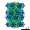
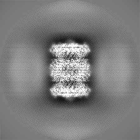
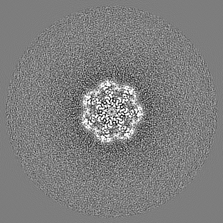
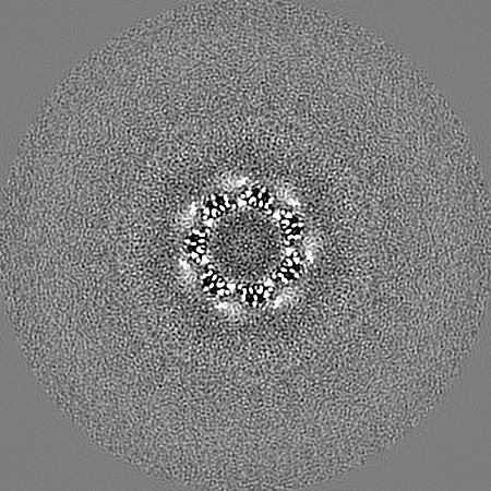
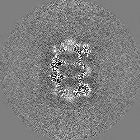
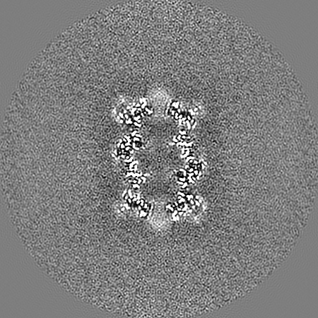
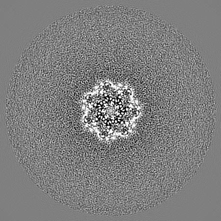
Journal: Structure / Year: 2020 Title: High-Throughput Cryo-EM Enabled by User-Free Preprocessing Routines. Authors: Yilai Li / Jennifer N Cash / John J G Tesmer / Michael A Cianfrocco / Abstract: Single-particle cryoelectron microscopy (cryo-EM) continues to grow into a mainstream structural biology technique. Recent developments in data collection strategies alongside new sample preparation ...Single-particle cryoelectron microscopy (cryo-EM) continues to grow into a mainstream structural biology technique. Recent developments in data collection strategies alongside new sample preparation devices herald a future where users will collect multiple datasets per microscope session. To make cryo-EM data processing more automatic and user-friendly, we have developed an automatic pipeline for cryo-EM data preprocessing and assessment using a combination of deep-learning and image-analysis tools. We have verified the performance of this pipeline on a number of datasets and extended its scope to include sample screening by the user-free assessment of the qualities of a series of datasets under different conditions. We propose that our workflow provides a decision-free solution for cryo-EM, making data preprocessing more generalized and robust in the high-throughput era as well as more convenient for users from a range of backgrounds.
History
Deposition
Feb 28, 2020
-
Header (metadata) release
Mar 11, 2020
-
Map release
Mar 11, 2020
-
Update
Jul 22, 2020
-
Current status
Jul 22, 2020
Processing site: RCSB / Status: Released
-
Structure visualization
Movie
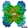
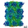
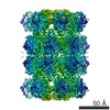
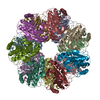
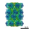
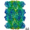
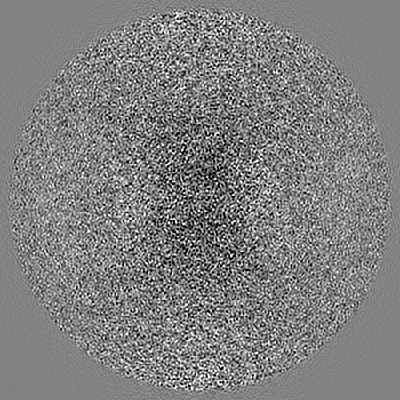
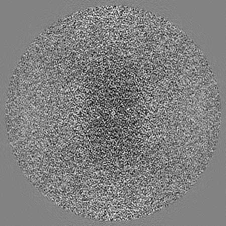
Surface view with section colored by density value
In the structure databanks used in Yorodumi, some data are registered as the other names, "COVID-19 virus" and "2019-nCoV". Here are the details of the virus and the list of structure data.
Jan 31, 2019. EMDB accession codes are about to change! (news from PDBe EMDB page)
EMDB accession codes are about to change! (news from PDBe EMDB page)
The allocation of 4 digits for EMDB accession codes will soon come to an end. Whilst these codes will remain in use, new EMDB accession codes will include an additional digit and will expand incrementally as the available range of codes is exhausted. The current 4-digit format prefixed with “EMD-” (i.e. EMD-XXXX) will advance to a 5-digit format (i.e. EMD-XXXXX), and so on. It is currently estimated that the 4-digit codes will be depleted around Spring 2019, at which point the 5-digit format will come into force.
The EM Navigator/Yorodumi systems omit the EMD- prefix.
Related info.:Q: What is EMD? / ID/Accession-code notation in Yorodumi/EM Navigator
Yorodumi is a browser for structure data from EMDB, PDB, SASBDB, etc.
This page is also the successor to EM Navigator detail page, and also detail information page/front-end page for Omokage search.
The word "yorodu" (or yorozu) is an old Japanese word meaning "ten thousand". "mi" (miru) is to see.
Related info.:EMDB / PDB / SASBDB / Comparison of 3 databanks / Yorodumi Search / Aug 31, 2016. New EM Navigator & Yorodumi / Yorodumi Papers / Jmol/JSmol / Function and homology information / Changes in new EM Navigator and Yorodumi
 Movie
Movie Controller

 Yorodumi
 Open data
 Basic information
 Map data
 Sample
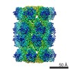
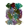
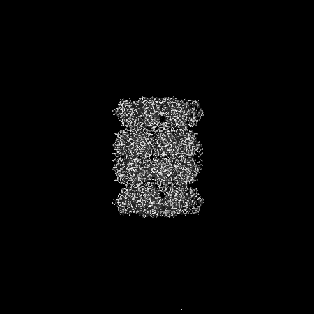
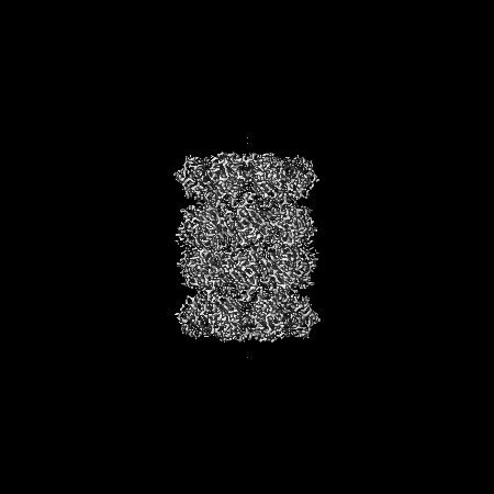
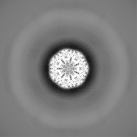
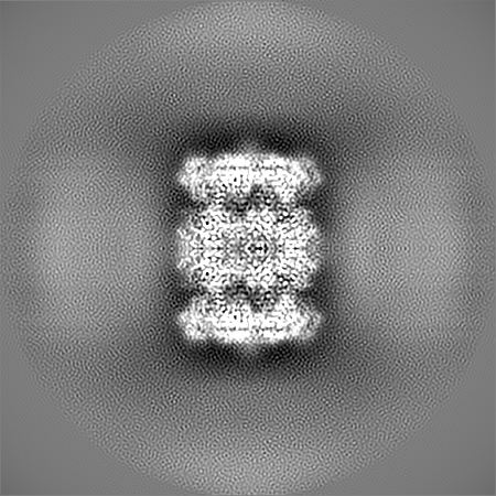
 Thermoplasma acidophilum (acidophilic)
 Authors
 United States, 2 items
  Citation
 Structure visualization
 Movie viewer
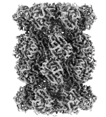
 Downloads & links
 emd_21491.png
 http://ftp.pdbj.org/pub/emdb/structures/EMD-21491
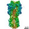
 Z (Sec.)
 Y (Row.)
 X (Col.)


 Sample components
 Processing
 Electron microscopy
 FIELD EMISSION GUN
