 Movie
Movie Controller

[English] 日本語
 Yorodumi
- EMDB-8999: Beam-tilt dependency of single-particle cryo-EM map quality: Expt... -
+ Open data

- Basic information
| Entry | Database: EMDB / ID: EMD-8999 | |||||||||
|---|---|---|---|---|---|---|---|---|---|---|
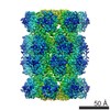
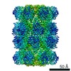
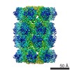
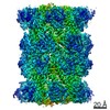
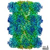
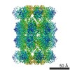
| Title | Beam-tilt dependency of single-particle cryo-EM map quality: Expt 2 at 0 mrad | |||||||||
 Map data Map data | Sharpened 0 beam tilt map in Experiment 2 | |||||||||
 Sample Sample |
| |||||||||
| Biological species |   Thermoplasma acidophilum (acidophilic) Thermoplasma acidophilum (acidophilic) | |||||||||
| Method | single particle reconstruction / cryo EM / Resolution: 2.74 Å | |||||||||
 Authors Authors | Cheng A / Eng ET / Alink L / Rice WJ / Jordan KD / Kim LY / Potter CS / Carragher B | |||||||||
 Citation Citation | Journal: J Struct Biol / Year: 2018 Title: High resolution single particle cryo-electron microscopy using beam-image shift. Authors: Anchi Cheng / Edward T Eng / Lambertus Alink / William J Rice / Kelsey D Jordan / Laura Y Kim / Clinton S Potter / Bridget Carragher /  Abstract: Automated data acquisition is used widely for single-particle reconstruction of three-dimensional (3D) volumes of biological complexes preserved in vitreous ice and imaged in a transmission electron ...Automated data acquisition is used widely for single-particle reconstruction of three-dimensional (3D) volumes of biological complexes preserved in vitreous ice and imaged in a transmission electron microscope. Automation has become integral to this method because of the very large number of particle images required in order to overcome the typically low signal-to-noise ratio of these images. For optimal efficiency, automated data acquisition software packages typically employ some beam-image shift targeting as this method is both fast and accurate (±0.1 µm). In contrast, using only stage movement, relocation to a targeted area under low-dose conditions can only be achieved in combination with multiple iterations or long relaxation times, both reducing efficiency. Nevertheless it is well known that applying beam-image shift induces beam-tilt and with it a potential structure phase error with a phase error π/4 the highest acceptable value. This theory has been used as an argument against beam-image shift for high resolution data collection. Nevertheless, in practice many small beam-image shift datasets have resulted in 3D reconstructions beyond the π/4 phase error limit. To address this apparent contradiction, we performed cryo-EM single-particle reconstructions on a T20S proteasome sample using applied beam-image shifts corresponding to beam tilts from 0 to 10 mrad. To evaluate the results we compared the FSC values, and examined the water density peaks in the 3D map. We conclude that the phase error does not limit the validity of the 3D reconstruction from single-particle averaging beyond the π/4 resolution limit. | |||||||||
| History |
|
- Structure visualization
| Movie |
 Movie viewer Movie viewer |
|---|---|
| Structure viewer | EM map: SurfViewMolmilJmol/JSmol |
| Supplemental images |

- Downloads & links
-EMDB archive
| Map data | emd_8999.map.gz | 59.5 MB | EMDB map data format | |
|---|---|---|---|---|
| Header (meta data) | emd-8999-v30.xmlemd-8999.xml | 12.1 KB 12.1 KB | Display Display | EMDB header |
| Images |  emd_8999.png emd_8999.png | 233.2 KB | ||
| Archive directory |  http://ftp.pdbj.org/pub/emdb/structures/EMD-8999ftp://ftp.pdbj.org/pub/emdb/structures/EMD-8999 http://ftp.pdbj.org/pub/emdb/structures/EMD-8999ftp://ftp.pdbj.org/pub/emdb/structures/EMD-8999 | HTTPS FTP |
-Related structure data
| Similar structure data |
|---|


-Links
| EMDB pages | EMDB (EBI/PDBe) / EMDataResource |
|---|
-Map
| File | Download / File: emd_8999.map.gz / Format: CCP4 / Size: 64 MB / Type: IMAGE STORED AS FLOATING POINT NUMBER (4 BYTES) | ||||||||||||||||||||||||||||||||||||||||||||||||||||||||||||
|---|---|---|---|---|---|---|---|---|---|---|---|---|---|---|---|---|---|---|---|---|---|---|---|---|---|---|---|---|---|---|---|---|---|---|---|---|---|---|---|---|---|---|---|---|---|---|---|---|---|---|---|---|---|---|---|---|---|---|---|---|---|
| Annotation | Sharpened 0 beam tilt map in Experiment 2 | ||||||||||||||||||||||||||||||||||||||||||||||||||||||||||||
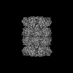
| Projections & slices | Image control
Images are generated by Spider. | ||||||||||||||||||||||||||||||||||||||||||||||||||||||||||||
| Voxel size | X=Y=Z: 1.0605 Å | ||||||||||||||||||||||||||||||||||||||||||||||||||||||||||||
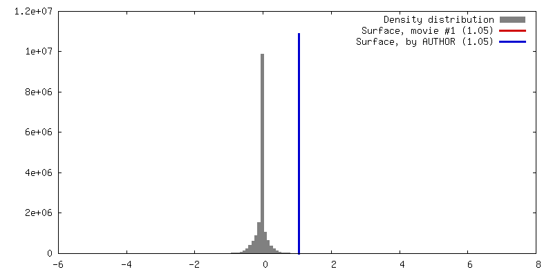
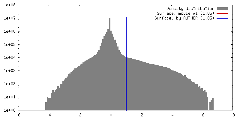
| Density |
| ||||||||||||||||||||||||||||||||||||||||||||||||||||||||||||
| Symmetry | Space group: 1 | ||||||||||||||||||||||||||||||||||||||||||||||||||||||||||||
| Details | EMDB XML:
CCP4 map header:
| ||||||||||||||||||||||||||||||||||||||||||||||||||||||||||||
 Z (Sec.)
 Y (Row.)
 X (Col.)


-Supplemental data
- Sample components
-Entire : Thermoplasma acidophilum 20S proteasome
| Entire | Name: Thermoplasma acidophilum 20S proteasome |
|---|---|
| Components |
|
-Supramolecule #1: Thermoplasma acidophilum 20S proteasome
| Supramolecule | Name: Thermoplasma acidophilum 20S proteasome / type: complex / ID: 1 / Parent: 0 |
|---|---|
| Source (natural) | Organism: Thermoplasma acidophilum (acidophilic) |
| Molecular weight | Theoretical: 700 KDa |
-Experimental details
-Structure determination
| Method | cryo EM |
|---|---|
 Processing Processing | single particle reconstruction |
| Aggregation state | particle |
-Sample preparation
| Concentration | 0.9 mg/mL | |||||||||
|---|---|---|---|---|---|---|---|---|---|---|
| Buffer | pH: 8 / Component:
| |||||||||
| Grid | Model: C-flat-1.2/1.3 / Material: COPPER / Mesh: 400 / Support film - Material: CARBON / Support film - topology: HOLEY / Pretreatment - Type: PLASMA CLEANING / Pretreatment - Atmosphere: OTHER | |||||||||
| Vitrification | Cryogen name: ETHANE / Chamber humidity: 75 % / Chamber temperature: 298 K / Instrument: GATAN CRYOPLUNGE 3 / Details: blotted for 2.5 s after 30 s wait time. |
- Electron microscopy
| Microscope | FEI TITAN KRIOS |
|---|---|
| Specialist optics | Energy filter - Name: GIF Quantum LS / Energy filter - Slit width: 15 eV |
| Details | 0,0 beam-image shift with Intentionally beam-shift pivot misalignment. |
| Image recording | Film or detector model: GATAN K2 SUMMIT (4k x 4k) / Detector mode: COUNTING / Digitization - Dimensions - Width: 3710 pixel / Digitization - Dimensions - Height: 3838 pixel / Digitization - Sampling interval: 5.0 µm / Digitization - Frames/image: 2-60 / Number grids imaged: 1 / Number real images: 545 / Average exposure time: 6.0 sec. / Average electron dose: 0.71 e/Å2 Details: of the 545 images, 158 images were removed from processing because of empty or high drift movie. |
| Electron beam | Acceleration voltage: 300 kV / Electron source:  FIELD EMISSION GUN FIELD EMISSION GUN |
| Electron optics | C2 aperture diameter: 70.0 µm / Illumination mode: FLOOD BEAM / Imaging mode: BRIGHT FIELD / Cs: 2.7 mm / Nominal defocus max: 2.0 µm / Nominal defocus min: 0.8 µm / Nominal magnification: 130000 |
| Sample stage | Specimen holder model: FEI TITAN KRIOS AUTOGRID HOLDER / Cooling holder cryogen: NITROGEN |
| Experimental equipment |  Model: Titan Krios / Image courtesy: FEI Company |
