Movie
Movie Controller

[English] 日本語
 Yorodumi
- EMDB-3109: Insight into the assembly of viruses with vertical single beta-ba... -
+ Open data

- Basic information
| Entry | Database: EMDB / ID: EMD-3109 | |||||||||
|---|---|---|---|---|---|---|---|---|---|---|
| Title | Insight into the assembly of viruses with vertical single beta-barrel major capsid proteins | |||||||||
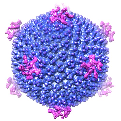
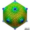
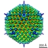
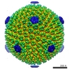
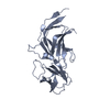
 Map data Map data | Reconstruction of Haloarcula hispanica icosahedral virus 2 (HHIV-2. The recommended contour level (threshold) suggested is for visualizing the capsid in Chimera. Lower thresholds are required for example to display the spikes. | |||||||||
 Sample Sample |
| |||||||||
 Keywords Keywords | archaeal virus | |||||||||
| Biological species |  Haloarcula hispanica icosahedral virus 2 Haloarcula hispanica icosahedral virus 2 | |||||||||
| Method | single particle reconstruction / cryo EM / Resolution: 13.0 Å | |||||||||
 Authors Authors | Gil-Carton D / Jaakkola ST / Charro D / Peralta B / Castano-Diez D / Oksanen HM / Bamford DH / Abrescia NG | |||||||||
 Citation Citation | Journal: Structure / Year: 2015 Title: Insight into the Assembly of Viruses with Vertical Single β-barrel Major Capsid Proteins. Authors: David Gil-Carton / Salla T Jaakkola / Diego Charro / Bibiana Peralta / Daniel Castaño-Díez / Hanna M Oksanen / Dennis H Bamford / Nicola G A Abrescia /    Abstract: Archaeal viruses constitute the least explored niche within the virosphere. Structure-based approaches have revealed close relationships between viruses infecting organisms from different domains of ...Archaeal viruses constitute the least explored niche within the virosphere. Structure-based approaches have revealed close relationships between viruses infecting organisms from different domains of life. Here, using biochemical and cryo-electron microscopy techniques, we solved the structure of euryarchaeal, halophilic, internal membrane-containing Haloarcula hispanica icosahedral virus 2 (HHIV-2). We show that the density of the two major capsid proteins (MCPs) recapitulates vertical single β-barrel proteins and that disulfide bridges stabilize the capsid. Below, ordered density is visible close to the membrane and at the five-fold vertices underneath the host-interacting vertex complex underpinning membrane-protein interactions. The HHIV-2 structure exemplifies the division of conserved architectural elements of a virion, such as the capsid, from those that evolve rapidly due to selective environmental pressure such as host-recognizing structures. We propose that in viruses with two vertical single β-barrel MCPs the vesicle is indispensable, and membrane-protein interactions serve as protein-railings for guiding the assembly. | |||||||||
| History |
|
- Structure visualization
| Movie |
 Movie viewer Movie viewer |
|---|---|
| Structure viewer | EM map: SurfViewMolmilJmol/JSmol |
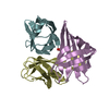
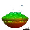
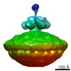
| Supplemental images |
- Downloads & links
-EMDB archive
| Map data | emd_3109.map.gz | 147.7 MB | EMDB map data format | |
|---|---|---|---|---|
| Header (meta data) | emd-3109-v30.xmlemd-3109.xml | 13.3 KB 13.3 KB | Display Display | EMDB header |
| Images | EMDB_figure_EMD-3109.tif | 465 KB | ||
| Archive directory |  http://ftp.pdbj.org/pub/emdb/structures/EMD-3109ftp://ftp.pdbj.org/pub/emdb/structures/EMD-3109 http://ftp.pdbj.org/pub/emdb/structures/EMD-3109ftp://ftp.pdbj.org/pub/emdb/structures/EMD-3109 | HTTPS FTP |
-Related structure data


-Links
| EMDB pages | EMDB (EBI/PDBe) / EMDataResource |
|---|
-Map
| File | Download / File: emd_3109.map.gz / Format: CCP4 / Size: 157 MB / Type: IMAGE STORED AS FLOATING POINT NUMBER (4 BYTES) | ||||||||||||||||||||||||||||||||||||||||||||||||||||||||||||
|---|---|---|---|---|---|---|---|---|---|---|---|---|---|---|---|---|---|---|---|---|---|---|---|---|---|---|---|---|---|---|---|---|---|---|---|---|---|---|---|---|---|---|---|---|---|---|---|---|---|---|---|---|---|---|---|---|---|---|---|---|---|
| Annotation | Reconstruction of Haloarcula hispanica icosahedral virus 2 (HHIV-2. The recommended contour level (threshold) suggested is for visualizing the capsid in Chimera. Lower thresholds are required for example to display the spikes. | ||||||||||||||||||||||||||||||||||||||||||||||||||||||||||||
| Projections & slices | Image control
Images are generated by Spider. | ||||||||||||||||||||||||||||||||||||||||||||||||||||||||||||
| Voxel size | X=Y=Z: 3.4 Å | ||||||||||||||||||||||||||||||||||||||||||||||||||||||||||||
| Density |
| ||||||||||||||||||||||||||||||||||||||||||||||||||||||||||||
| Symmetry | Space group: 1 | ||||||||||||||||||||||||||||||||||||||||||||||||||||||||||||
| Details | EMDB XML:
CCP4 map header:
| ||||||||||||||||||||||||||||||||||||||||||||||||||||||||||||
 Z (Sec.)
 Y (Row.)
 X (Col.)


-Supplemental data
- Sample components
-Entire : Haloarcula hispanica icosahedral virus 2 (HHIV-2)
| Entire | Name: Haloarcula hispanica icosahedral virus 2 (HHIV-2) |
|---|---|
| Components |
|
-Supramolecule #1000: Haloarcula hispanica icosahedral virus 2 (HHIV-2)
| Supramolecule | Name: Haloarcula hispanica icosahedral virus 2 (HHIV-2) / type: sample / ID: 1000 / Oligomeric state: Icosahedral Virus / Number unique components: 1 |
|---|
-Supramolecule #1: Haloarcula hispanica icosahedral virus 2
| Supramolecule | Name: Haloarcula hispanica icosahedral virus 2 / type: virus / ID: 1 / Details: HHIV-2 is a lipid-containing virus / NCBI-ID: 1154689 / Sci species name: Haloarcula hispanica icosahedral virus 2 / Virus type: VIRION / Virus isolate: SPECIES / Virus enveloped: No / Virus empty: No |
|---|---|
| Host (natural) | Organism:  Haloarcula hispanica (Halophile) / synonym: ARCHAEA Haloarcula hispanica (Halophile) / synonym: ARCHAEA |
| Virus shell | Shell ID: 1 / Name: VP4-VP7 / Diameter: 740 Å / T number (triangulation number): 28 |
-Experimental details
-Structure determination
| Method | cryo EM |
|---|---|
 Processing Processing | single particle reconstruction |
| Aggregation state | particle |
-Sample preparation
| Concentration | 0.9 mg/mL |
|---|---|
| Buffer | pH: 7.2 Details: 20 mM Tris-HCl [pH 7.2], 20 mM MgCl2, 10 mM CaCl2, and 0.5 M NaCl |
| Grid | Details: 200-mesh Quantifoil R 2/1 holey-carbon grids |
| Vitrification | Cryogen name: ETHANE / Chamber humidity: 95 % / Chamber temperature: 100 K / Instrument: FEI VITROBOT MARK II / Method: Blot for 3 seconds before plunging |
- Electron microscopy
| Microscope | JEOL 2200FSC |
|---|---|
| Temperature | Min: 80 K / Max: 103 K / Average: 99 K |
| Alignment procedure | Legacy - Astigmatism: Objective lens astigmatism was corrected at 100,000 times magnification |
| Specialist optics | Energy filter - Name: Omega / Energy filter - Lower energy threshold: 0.0 eV / Energy filter - Upper energy threshold: 15.0 eV |
| Date | Oct 27, 2014 |
| Image recording | Category: CCD / Film or detector model: GATAN ULTRASCAN 4000 (4k x 4k) / Digitization - Sampling interval: 15 µm / Number real images: 900 / Average electron dose: 10 e/Å2 / Bits/pixel: 32 |
| Electron beam | Acceleration voltage: 200 kV / Electron source:  FIELD EMISSION GUN FIELD EMISSION GUN |
| Electron optics | Calibrated magnification: 90200 / Illumination mode: FLOOD BEAM / Imaging mode: BRIGHT FIELD / Cs: 2 mm / Nominal defocus max: 2.8 µm / Nominal defocus min: 0.8 µm / Nominal magnification: 60000 |
| Sample stage | Specimen holder model: GATAN LIQUID NITROGEN |
-Image processing
| CTF correction | Details: micrograph |
|---|---|
| Final reconstruction | Applied symmetry - Point group: I (icosahedral) / Algorithm: OTHER / Resolution.type: BY AUTHOR / Resolution: 13.0 Å / Resolution method: OTHER / Software - Name: XMIPP / Details: 11 Ang FSC at 0.143 cut-off / Number images used: 4875 |
-Atomic model buiding 1
| Initial model | PDB ID: |
|---|---|
| Software | Name: COOT |
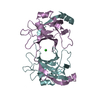
| Details | We used the VP17 and VP16 pdbs previously fitted into the P23-77 cryo-EM maps by Rissanen et al (2013) Structure. We selected from the above fitting as rigid body a VP17 with two adjacent VP16. Then, we fitted as rigid body the VP16-VP17-VP16 oligomer in our map into capsomer 5 (please see our publication Figure 4D). |
| Refinement | Space: REAL / Protocol: RIGID BODY FIT |
-Atomic model buiding 2
| Initial model | PDB ID: |
|---|---|
| Software | Name: COOT |
| Details | We used the VP17 and VP16 pdbs previously fitted into the P23-77 cryo-EM maps by Rissanen et al (2013) Structure. We selected from the above fitting as rigid body a VP17 with two adjacent VP16. Then, we fitted as rigid body the VP16-VP17-VP16 oligomer in our map into capsomer 5 using the COOT command 'Rigid body fit zone' (please see our publication Figure 4D). |
| Refinement | Space: REAL / Protocol: RIGID BODY FIT |
-Atomic model buiding 3
| Initial model | PDB ID: Chain - Chain ID: Q |
|---|---|
| Software | Name: COOT |
| Details | We first generated the pentameric structure correspondingto the STIV map - Veesler et al (2013) PNAS- and then used the pentamer for rigid-body fitting using the COOT command 'Rigid body fit zone' in our cryo-EM density (please see Figure 7). |
| Refinement | Space: REAL / Protocol: RIGID BODY FIT |

-Atomic model buiding 4
| Initial model | PDB ID: |
|---|---|
| Software | Name: COOT |
| Details | The trimer was manually fitted into density using COOT |
| Refinement | Space: REAL / Protocol: RIGID BODY FIT |

-Atomic model buiding 5
| Initial model | PDB ID: |
|---|---|
| Software | Name: COOT |
| Details | The trimer was manually fitted into density using COOT. |
| Refinement | Space: REAL / Protocol: RIGID BODY FIT |