Movie
Movie Controller

[English] 日本語
 Yorodumi
- EMDB-2063: A 3-D cryo-electron microscopy structure of bacteriophage phi92 capsid -
+ Open data

- Basic information
| Entry | Database: EMDB / ID: EMD-2063 | |||||||||
|---|---|---|---|---|---|---|---|---|---|---|
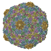
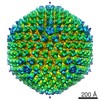
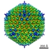
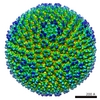
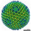
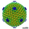
| Title | A 3-D cryo-electron microscopy structure of bacteriophage phi92 capsid | |||||||||
 Map data Map data | Reconstruction ofbacteriophage phi92 capsid | |||||||||
 Sample Sample |
| |||||||||
 Keywords Keywords | Bacteriophage / phi92 / capsid / single particle / cryo-electron / reconstruction | |||||||||
| Biological species |  Staphylococcus phage 92 (virus) Staphylococcus phage 92 (virus) | |||||||||
| Method | single particle reconstruction / cryo EM / negative staining / Resolution: 19.0 Å | |||||||||
 Authors Authors | Browning C / Nazarov S / Bowman V / Leiman P | |||||||||
 Citation Citation | Journal: J Virol / Year: 2012 Title: A multivalent adsorption apparatus explains the broad host range of phage phi92: a comprehensive genomic and structural analysis. Authors: David Schwarzer / Falk F R Buettner / Christopher Browning / Sergey Nazarov / Wolfgang Rabsch / Andrea Bethe / Astrid Oberbeck / Valorie D Bowman / Katharina Stummeyer / Martina Mühlenhoff ...Authors: David Schwarzer / Falk F R Buettner / Christopher Browning / Sergey Nazarov / Wolfgang Rabsch / Andrea Bethe / Astrid Oberbeck / Valorie D Bowman / Katharina Stummeyer / Martina Mühlenhoff / Petr G Leiman / Rita Gerardy-Schahn /  Abstract: Bacteriophage phi92 is a large, lytic myovirus isolated in 1983 from pathogenic Escherichia coli strains that carry a polysialic acid capsule. Here we report the genome organization of phi92, the ...Bacteriophage phi92 is a large, lytic myovirus isolated in 1983 from pathogenic Escherichia coli strains that carry a polysialic acid capsule. Here we report the genome organization of phi92, the cryoelectron microscopy reconstruction of its virion, and the reinvestigation of its host specificity. The genome consists of a linear, double-stranded 148,612-bp DNA sequence containing 248 potential open reading frames and 11 putative tRNA genes. Orthologs were found for 130 of the predicted proteins. Most of the virion proteins showed significant sequence similarities to proteins of myoviruses rv5 and PVP-SE1, indicating that phi92 is a new member of the novel genus of rv5-like phages. Reinvestigation of phi92 host specificity showed that the host range is not limited to polysialic acid-encapsulated Escherichia coli but includes most laboratory strains of Escherichia coli and many Salmonella strains. Structure analysis of the phi92 virion demonstrated the presence of four different types of tail fibers and/or tailspikes, which enable the phage to use attachment sites on encapsulated and nonencapsulated bacteria. With this report, we provide the first detailed description of a multivalent, multispecies phage armed with a host cell adsorption apparatus resembling a nanosized Swiss army knife. The genome, structure, and, in particular, the organization of the baseplate of phi92 demonstrate how a bacteriophage can evolve into a multi-pathogen-killing agent. | |||||||||
| History |
|
- Structure visualization
| Movie |
 Movie viewer Movie viewer |
|---|---|
| Structure viewer | EM map: SurfViewMolmilJmol/JSmol |
| Supplemental images |

- Downloads & links
-EMDB archive
| Map data | emd_2063.map.gz | 105.2 MB | EMDB map data format | |
|---|---|---|---|---|
| Header (meta data) | emd-2063-v30.xmlemd-2063.xml | 9.5 KB 9.5 KB | Display Display | EMDB header |
| Images | 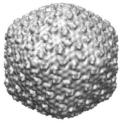 2063.png 2063.png | 118.3 KB | ||
| Archive directory |  http://ftp.pdbj.org/pub/emdb/structures/EMD-2063ftp://ftp.pdbj.org/pub/emdb/structures/EMD-2063 http://ftp.pdbj.org/pub/emdb/structures/EMD-2063ftp://ftp.pdbj.org/pub/emdb/structures/EMD-2063 | HTTPS FTP |
-Related structure data


-Links
| EMDB pages | EMDB (EBI/PDBe) / EMDataResource |
|---|
-Map
| File | Download / File: emd_2063.map.gz / Format: CCP4 / Size: 210.9 MB / Type: IMAGE STORED AS FLOATING POINT NUMBER (4 BYTES) | ||||||||||||||||||||||||||||||||||||||||||||||||||||||||||||||||||||
|---|---|---|---|---|---|---|---|---|---|---|---|---|---|---|---|---|---|---|---|---|---|---|---|---|---|---|---|---|---|---|---|---|---|---|---|---|---|---|---|---|---|---|---|---|---|---|---|---|---|---|---|---|---|---|---|---|---|---|---|---|---|---|---|---|---|---|---|---|---|
| Annotation | Reconstruction ofbacteriophage phi92 capsid | ||||||||||||||||||||||||||||||||||||||||||||||||||||||||||||||||||||
| Projections & slices | Image control
Images are generated by Spider. | ||||||||||||||||||||||||||||||||||||||||||||||||||||||||||||||||||||
| Voxel size | X=Y=Z: 2.81 Å | ||||||||||||||||||||||||||||||||||||||||||||||||||||||||||||||||||||
| Density |
| ||||||||||||||||||||||||||||||||||||||||||||||||||||||||||||||||||||
| Symmetry | Space group: 1 | ||||||||||||||||||||||||||||||||||||||||||||||||||||||||||||||||||||
| Details | EMDB XML:
CCP4 map header:
| ||||||||||||||||||||||||||||||||||||||||||||||||||||||||||||||||||||
 Z (Sec.)
 Y (Row.)
 X (Col.)


-Supplemental data
- Sample components
-Entire : Bacteriophage phi92
| Entire | Name: Bacteriophage phi92 (virus) |
|---|---|
| Components |
|
-Supramolecule #1000: Bacteriophage phi92
| Supramolecule | Name: Bacteriophage phi92 / type: sample / ID: 1000 / Number unique components: 1 |
|---|
-Supramolecule #1: Staphylococcus phage 92
| Supramolecule | Name: Staphylococcus phage 92 / type: virus / ID: 1 / Name.synonym: bacteriophage phi92 / Details: Bacteriophage phi92 is a large lytic myovirus / NCBI-ID: 320849 / Sci species name: Staphylococcus phage 92 / Virus type: VIRION / Virus isolate: STRAIN / Virus enveloped: No / Virus empty: No / Syn species name: bacteriophage phi92 |
|---|---|
| Host (natural) | Organism:  |
| Virus shell | Shell ID: 1 / T number (triangulation number): 13 |
-Experimental details
-Structure determination
| Method | negative staining, cryo EM |
|---|---|
 Processing Processing | single particle reconstruction |
| Aggregation state | particle |
-Sample preparation
| Buffer | Details: 50mM TrisCl pH 7.5, 100mM NaCl, 8mM MgSO4 |
|---|---|
| Staining | Type: NEGATIVE / Details: vitrification in liquid ethane |
| Grid | Details: holey carbon grid |
| Vitrification | Cryogen name: ETHANE / Chamber humidity: 100 % / Chamber temperature: 113 K / Instrument: HOMEMADE PLUNGER |
- Electron microscopy
| Microscope | FEI/PHILIPS CM300FEG/T |
|---|---|
| Date | May 30, 2007 |
| Image recording | Category: FILM / Film or detector model: KODAK SO-163 FILM / Digitization - Scanner: ZEISS SCAI / Digitization - Sampling interval: 7 µm / Number real images: 38 / Average electron dose: 20 e/Å2 |
| Electron beam | Acceleration voltage: 300 kV / Electron source:  FIELD EMISSION GUN FIELD EMISSION GUN |
| Electron optics | Illumination mode: FLOOD BEAM / Imaging mode: BRIGHT FIELD / Cs: 2.0 mm / Nominal defocus max: 3.5 µm / Nominal defocus min: 2.0 µm / Nominal magnification: 33000 |
| Sample stage | Specimen holder model: PHILIPS ROTATION HOLDER |
-Image processing
| Details | The icosahedral reconstruction the phi92 capsid was calculated from scratch. The initial model was constructed with a small subset of initial data. This model was then subjected to several rounds of refinement until convergence. |
|---|---|
| CTF correction | Details: Each Micrograph |
| Final reconstruction | Applied symmetry - Point group: I (icosahedral) / Algorithm: OTHER / Resolution.type: BY AUTHOR / Resolution: 19.0 Å / Resolution method: FSC 0.5 CUT-OFF / Software - Name: EMAN / Number images used: 1137 |
| Final two d classification | Number classes: 10 |
-Atomic model buiding 1
| Initial model | PDB ID: |
|---|---|
| Software | Name: Chimera |
| Refinement | Space: REAL |