 Movie
Movie Controller

[English] 日本語
 Yorodumi
- PDB-3j31: Life in the extremes: atomic structure of Sulfolobus Turreted Ico... -
+ Open data

- Basic information
| Entry | Database: PDB / ID: 3j31 | ||||||
|---|---|---|---|---|---|---|---|
| Title | Life in the extremes: atomic structure of Sulfolobus Turreted Icosahedral Virus | ||||||
 Components Components |
| ||||||
 Keywords Keywords | VIRUS / Virus assembly / Evolution / Archaea | ||||||
| Function / homology |  Function and homology information Function and homology information | ||||||
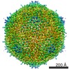
| Biological species |    Sulfolobus turreted icosahedral virus Sulfolobus turreted icosahedral virus | ||||||
| Method | ELECTRON MICROSCOPY / single particle reconstruction / cryo EM / Resolution: 4.5 Å | ||||||
 Authors Authors | Veesler, D. / Ng, T.S. / Sendamarai, A.K. / Eilers, B.J. / Lawrence, C.M. / Lok, S.M. / Young, M.J. / Johnson, J.E. / Fu, C.-Y. | ||||||
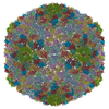
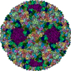
 Citation Citation | Journal: Proc Natl Acad Sci U S A / Year: 2013 Title: Atomic structure of the 75 MDa extremophile Sulfolobus turreted icosahedral virus determined by CryoEM and X-ray crystallography. Authors: David Veesler / Thiam-Seng Ng / Anoop K Sendamarai / Brian J Eilers / C Martin Lawrence / Shee-Mei Lok / Mark J Young / John E Johnson / Chi-yu Fu /  Abstract: Sulfolobus turreted icosahedral virus (STIV) was isolated in acidic hot springs where it infects the archeon Sulfolobus solfataricus. We determined the STIV structure using near-atomic resolution ...Sulfolobus turreted icosahedral virus (STIV) was isolated in acidic hot springs where it infects the archeon Sulfolobus solfataricus. We determined the STIV structure using near-atomic resolution electron microscopy and X-ray crystallography allowing tracing of structural polypeptide chains and visualization of transmembrane proteins embedded in the viral membrane. We propose that the vertex complexes orchestrate virion assembly by coordinating interactions of the membrane and various protein components involved. STIV shares the same coat subunit and penton base protein folds as some eukaryotic and bacterial viruses, suggesting that they derive from a common ancestor predating the divergence of the three kingdoms of life. One architectural motif (β-jelly roll fold) forms virtually the entire capsid (distributed in three different gene products), indicating that a single ancestral protein module may have been at the origin of its evolution. | ||||||
| History |
|
- Structure visualization
| Movie |
Movie viewer |
|---|---|
| Structure viewer | Molecule: MolmilJmol/JSmol |
- Downloads & links
-Download
| PDBx/mmCIF format | 3j31.cif.gz | 1 MB | Display | PDBx/mmCIF format |
|---|---|---|---|---|
| PDB format | pdb3j31.ent.gz | 876 KB | Display | PDB format |
| PDBx/mmJSON format | 3j31.json.gz | Tree view | PDBx/mmJSON format | |
| Others |  Other downloads Other downloads |
-Validation report
| Arichive directory | https://data.pdbj.org/pub/pdb/validation_reports/j3/3j31ftp://data.pdbj.org/pub/pdb/validation_reports/j3/3j31 | HTTPS FTP |
|---|
-Related structure data
| Related structure data | 5584MC 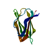 4il7C M: map data used to model this data C: citing same article ( |
|---|---|
| Similar structure data |


-Links
 PDBj
PDBj- Assembly
| Deposited unit | 
|
|---|---|
| 1 | x 60
|
| 2 |
|
| 3 | x 5
|
| 4 | x 6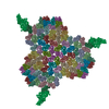
|
| 5 | 
|
| Symmetry | Point symmetry: (Schoenflies symbol: I (icosahedral)) |
-Components
| #1: Protein | Mass: 24439.609 Da / Num. of mol.: 1 / Source method: isolated from a natural source / Source: (natural) Sulfolobus turreted icosahedral virus / References: UniProt: Q6Q0L4 | ||||
|---|---|---|---|---|---|
| #2: Protein/peptide | Mass: 1294.587 Da / Num. of mol.: 1 / Fragment: SEE REMARK 999 / Source method: isolated from a natural source / Source: (natural) Sulfolobus turreted icosahedral virus | ||||
| #3: Protein | Mass: 37858.805 Da / Num. of mol.: 15 / Source method: isolated from a natural source / Source: (natural) Sulfolobus turreted icosahedral virus / References: UniProt: Q6Q0J0#4: Protein | | Mass: 41706.789 Da / Num. of mol.: 1 / Source method: isolated from a natural source / Source: (natural) Sulfolobus turreted icosahedral virus / References: UniProt: Q6Q0L3Sequence details | CHAIN R CORRESPONDS TO A PORTION OF THE SEQUENCE ...CHAIN R CORRESPOND | |
-Experimental details
-Experiment
| Experiment | Method: ELECTRON MICROSCOPY |
|---|---|
| EM experiment | Aggregation state: PARTICLE / 3D reconstruction method: single particle reconstruction |
- Sample preparation
| Component | Name: Mature Sulfolobus Turreted Icosahedral Virus / Type: VIRUS |
|---|---|
| Molecular weight | Value: 75 MDa / Experimental value: NO |
| Details of virus | Empty: NO / Enveloped: YES / Host category: ARCHAEA / Isolate: STRAIN / Type: VIRION |
| Natural host | Organism: Sulfolobus solfataricus |
| Buffer solution | Name: 23 mM KH2PO4, 19 mM (NH4)2SO4, 1 mM MgSO4, 2 mM CaCl2 / pH: 3.5 Details: 23 mM KH2PO4, 19 mM (NH4)2SO4, 1 mM MgSO4, 2 mM CaCl2 |
| Specimen | Embedding applied: NO / Shadowing applied: NO / Staining applied: NO / Vitrification applied: YES |
| Specimen support | Details: glow-discharged C-flat holey carbon grids (CF-2/2-4C, Protochips) |
| Vitrification | Instrument: HOMEMADE PLUNGER / Cryogen name: ETHANE / Temp: 94 K Details: Vitrification carried out at room temperature using liquid ethane and a homemade plunger |
- Electron microscopy imaging
| Experimental equipment |  Model: Titan Krios / Image courtesy: FEI Company |
|---|---|
| Microscopy | Model: FEI TITAN KRIOS / Date: Mar 30, 2011 / Details: Nanoprobe mode was used. |
| Electron gun | Electron source:  FIELD EMISSION GUN / Accelerating voltage: 300 kV / Illumination mode: FLOOD BEAM FIELD EMISSION GUN / Accelerating voltage: 300 kV / Illumination mode: FLOOD BEAM |
| Electron lens | Mode: BRIGHT FIELD / Calibrated magnification: 102189 X / Nominal defocus max: 2500 nm / Nominal defocus min: 800 nm / Cs: 2.7 mm Astigmatism: Objective lens astigmatism was corrected at 103000 times magnification Camera length: 0 mm |
| Specimen holder | Specimen holder model: FEI TITAN KRIOS AUTOGRID HOLDER / Specimen holder type: Nitrogen cooled / Temperature: 90 K / Tilt angle max: 0 ° / Tilt angle min: 0 ° |
| Image recording | Electron dose: 18 e/Å2 / Film or detector model: FEI FALCON I (4k x 4k) / Details: Non-backthinned chip |
| Image scans | Num. digital images: 878 |
| Radiation | Protocol: SINGLE WAVELENGTH / Monochromatic (M) / Laue (L): M / Scattering type: x-ray |
| Radiation wavelength | Relative weight: 1 |
- Processing
| EM software |
| |||||||||||||||||||||
|---|---|---|---|---|---|---|---|---|---|---|---|---|---|---|---|---|---|---|---|---|---|---|
| CTF correction | Details: Each particle | |||||||||||||||||||||
| Symmetry | Point symmetry: I (icosahedral) | |||||||||||||||||||||
| 3D reconstruction | Method: Projection matching / Resolution: 4.5 Å / Resolution method: FSC 0.143 CUT-OFF / Num. of particles: 8903 / Actual pixel size: 1.37 Å / Magnification calibration: Fitting of atomic coordinates Details: The final reconstruction was sharpened with a negative temperature factor of 400A2 (Single particle--Applied symmetry: I) Resolution method was FSC at 0.143 cut-off. The reported resolution ...Details: The final reconstruction was sharpened with a negative temperature factor of 400A2 (Single particle--Applied symmetry: I) Resolution method was FSC at 0.143 cut-off. The reported resolution is for the entire reconstruction. The resolution of the coat subunit region (B345) is estimated to 3.9 A using the same criterion. Symmetry type: POINT | |||||||||||||||||||||
| Atomic model building |
| |||||||||||||||||||||
| Atomic model building |
| |||||||||||||||||||||
| Refinement step | Cycle: LAST
|

