 Movie
Movie Controller

[English] 日本語
 Yorodumi
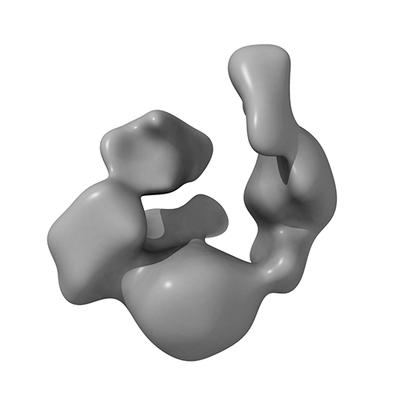
- EMDB-30386: Negative-stain EM 3D reconstruction of UGGT with the Fab of monoc... -
+ Open data

- Basic information
| Entry | Database: EMDB / ID: EMD-30386 | |||||||||
|---|---|---|---|---|---|---|---|---|---|---|
| Title | Negative-stain EM 3D reconstruction of UGGT with the Fab of monoclonal antibody directed against the Trx4 domain. | |||||||||
 Map data Map data | Negative-stain EM 3D reconstruction of UGGT with the Fab of monoclonal antibody | |||||||||
 Sample Sample |
| |||||||||
| Biological species |   Thermomyces dupontii (fungus) Thermomyces dupontii (fungus) | |||||||||
| Method | single particle reconstruction / negative staining / Resolution: 22.9 Å | |||||||||
 Authors Authors | Satoh T / Song C / Murata K / Kato K | |||||||||
 Citation Citation | Journal: Sci Rep / Year: 2017 Title: Visualisation of a flexible modular structure of the ER folding-sensor enzyme UGGT. Authors: Tadashi Satoh / Chihong Song / Tong Zhu / Takayasu Toshimori / Kazuyoshi Murata / Yugo Hayashi / Hironari Kamikubo / Takayuki Uchihashi / Koichi Kato /  Abstract: In the endoplasmic reticulum (ER), a protein quality control system facilitates the efficient folding of newly synthesised proteins. In this system, a series of N-linked glycan intermediates ...In the endoplasmic reticulum (ER), a protein quality control system facilitates the efficient folding of newly synthesised proteins. In this system, a series of N-linked glycan intermediates displayed on the protein surface serve as quality tags. The ER folding-sensor enzyme UDP-glucose:glycoprotein glucosyltransferase (UGGT) acts as a gatekeeper in the ER quality control system by specifically catalysing monoglucosylation onto incompletely folded glycoproteins, thereby enabling them to interact with lectin-chaperone complexes. Here we characterise the dynamic structure of this enzyme. Our crystallographic data demonstrate that the sensor region is composed of four thioredoxin-like domains followed by a β-rich domain, which are arranged into a C-shaped structure with a large central cavity, while the C-terminal catalytic domain undergoes a ligand-dependent conformational alteration. Furthermore, small-angle X-ray scattering, cryo-electron microscopy and high-speed atomic force microscopy have demonstrated that UGGT has a flexible modular structure in which the smaller catalytic domain is tethered to the larger folding-sensor region with variable spatial arrangements. These findings provide structural insights into the working mechanism whereby UGGT operates as a folding-sensor against a variety of glycoprotein substrates through its flexible modular structure possessing extended hydrophobic surfaces for the recognition of unfolded substrates. | |||||||||
| History |
|
- Structure visualization
| Movie |
 Movie viewer Movie viewer |
|---|---|
| Structure viewer | EM map: SurfViewMolmilJmol/JSmol |
| Supplemental images |
- Downloads & links
-EMDB archive
| Map data | emd_30386.map.gz | 27.8 MB | EMDB map data format | |
|---|---|---|---|---|
| Header (meta data) | emd-30386-v30.xmlemd-30386.xml | 7.6 KB 7.6 KB | Display Display | EMDB header |
| Images |  emd_30386.png emd_30386.png | 35.8 KB | ||
| Archive directory |  http://ftp.pdbj.org/pub/emdb/structures/EMD-30386ftp://ftp.pdbj.org/pub/emdb/structures/EMD-30386 http://ftp.pdbj.org/pub/emdb/structures/EMD-30386ftp://ftp.pdbj.org/pub/emdb/structures/EMD-30386 | HTTPS FTP |
-Validation report
| Summary document | emd_30386_validation.pdf.gz | 78.4 KB | Display | EMDB validaton report |
|---|---|---|---|---|
| Full document | emd_30386_full_validation.pdf.gz | 77.5 KB | Display | |
| Data in XML | emd_30386_validation.xml.gz | 496 B | Display | |
| Arichive directory | https://ftp.pdbj.org/pub/emdb/validation_reports/EMD-30386ftp://ftp.pdbj.org/pub/emdb/validation_reports/EMD-30386 | HTTPS FTP |
-Related structure data


-Links
| EMDB pages | EMDB (EBI/PDBe) / EMDataResource |
|---|
-Map
| File | Download / File: emd_30386.map.gz / Format: CCP4 / Size: 83.7 MB / Type: IMAGE STORED AS FLOATING POINT NUMBER (4 BYTES) | ||||||||||||||||||||||||||||||||||||||||||||||||||||||||||||||||||||
|---|---|---|---|---|---|---|---|---|---|---|---|---|---|---|---|---|---|---|---|---|---|---|---|---|---|---|---|---|---|---|---|---|---|---|---|---|---|---|---|---|---|---|---|---|---|---|---|---|---|---|---|---|---|---|---|---|---|---|---|---|---|---|---|---|---|---|---|---|---|
| Annotation | Negative-stain EM 3D reconstruction of UGGT with the Fab of monoclonal antibody | ||||||||||||||||||||||||||||||||||||||||||||||||||||||||||||||||||||
| Projections & slices | Image control
Images are generated by Spider. | ||||||||||||||||||||||||||||||||||||||||||||||||||||||||||||||||||||
| Voxel size | X=Y=Z: 0.98 Å | ||||||||||||||||||||||||||||||||||||||||||||||||||||||||||||||||||||
| Density |
| ||||||||||||||||||||||||||||||||||||||||||||||||||||||||||||||||||||
| Symmetry | Space group: 1 | ||||||||||||||||||||||||||||||||||||||||||||||||||||||||||||||||||||
| Details | EMDB XML:
CCP4 map header:
| ||||||||||||||||||||||||||||||||||||||||||||||||||||||||||||||||||||
 Z (Sec.)
 Y (Row.)
 X (Col.)


-Supplemental data
- Sample components
-Entire : UGGT with the Fab of monoclonal antibody directed against the Trx...
| Entire | Name: UGGT with the Fab of monoclonal antibody directed against the Trx4 domain. |
|---|---|
| Components |
|
-Supramolecule #1: UGGT with the Fab of monoclonal antibody directed against the Trx...
| Supramolecule | Name: UGGT with the Fab of monoclonal antibody directed against the Trx4 domain. type: complex / ID: 1 / Parent: 0 |
|---|---|
| Source (natural) | Organism: Thermomyces dupontii (fungus) |
| Recombinant expression | Organism:  Tobacco etch virus Tobacco etch virus |
-Experimental details
-Structure determination
| Method | negative staining |
|---|---|
 Processing Processing | single particle reconstruction |
| Aggregation state | particle |
-Sample preparation
| Buffer | pH: 7.7 |
|---|---|
| Staining | Type: NEGATIVE / Material: uranyl acetate |
- Electron microscopy
| Microscope | JEOL 2200FS |
|---|---|
| Image recording | Film or detector model: DIRECT ELECTRON DE-20 (5k x 3k) / Average electron dose: 40.0 e/Å2 |
| Electron beam | Acceleration voltage: 200 kV / Electron source:  FIELD EMISSION GUN FIELD EMISSION GUN |
| Electron optics | Illumination mode: FLOOD BEAM / Imaging mode: BRIGHT FIELD |
-Image processing
| Final reconstruction | Resolution.type: BY AUTHOR / Resolution: 22.9 Å / Resolution method: FSC 0.143 CUT-OFF / Number images used: 2251 |
|---|---|
| Initial angle assignment | Type: ANGULAR RECONSTITUTION |
| Final angle assignment | Type: ANGULAR RECONSTITUTION |
