 Movie
Movie Controller


+ Open data

- Basic information
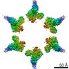
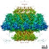
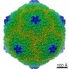
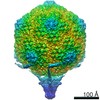
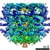
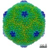
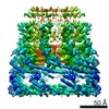
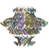
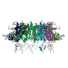
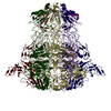
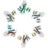
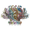
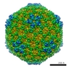
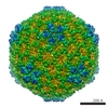
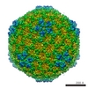
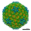
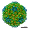
| Entry | Database: EMDB / ID: EMD-30144 | |||||||||
|---|---|---|---|---|---|---|---|---|---|---|
| Title | Phage t7 cpasid-II mismatch | |||||||||
 Map data Map data | ||||||||||
 Sample Sample |
| |||||||||
| Biological species |   Escherichia phage T7 (virus) Escherichia phage T7 (virus) | |||||||||
| Method | single particle reconstruction / cryo EM / Resolution: 5.9 Å | |||||||||
 Authors Authors | Chen WY / Xiao H | |||||||||
| Funding support | 1 items
| |||||||||
 Citation Citation | Journal: Protein Cell / Year: 2020 Title: Structural changes of a bacteriophage upon DNA packaging and maturation. Authors: Wenyuan Chen / Hao Xiao / Xurong Wang / Shuanglin Song / Zhen Han / Xiaowu Li / Fan Yang / Li Wang / Jingdong Song / Hongrong Liu / Lingpeng Cheng /  | |||||||||
| History |
|
- Structure visualization
| Movie |
 Movie viewer Movie viewer |
|---|---|
| Structure viewer | EM map: SurfViewMolmilJmol/JSmol |
| Supplemental images |

- Downloads & links
-EMDB archive
| Map data | emd_30144.map.gz | 190.4 MB | EMDB map data format | |
|---|---|---|---|---|
| Header (meta data) | emd-30144-v30.xmlemd-30144.xml | 7.5 KB 7.5 KB | Display Display | EMDB header |
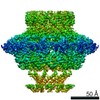
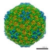
| Images | 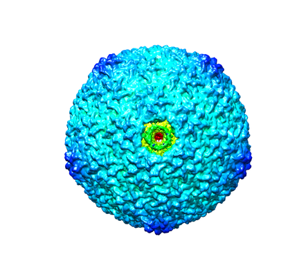 emd_30144.png emd_30144.png | 131.3 KB | ||
| Archive directory |  http://ftp.pdbj.org/pub/emdb/structures/EMD-30144ftp://ftp.pdbj.org/pub/emdb/structures/EMD-30144 http://ftp.pdbj.org/pub/emdb/structures/EMD-30144ftp://ftp.pdbj.org/pub/emdb/structures/EMD-30144 | HTTPS FTP |
-Validation report
| Summary document | emd_30144_validation.pdf.gz | 383.3 KB | Display | EMDB validaton report |
|---|---|---|---|---|
| Full document | emd_30144_full_validation.pdf.gz | 382.9 KB | Display | |
| Data in XML | emd_30144_validation.xml.gz | 7.6 KB | Display | |
| Arichive directory | https://ftp.pdbj.org/pub/emdb/validation_reports/EMD-30144ftp://ftp.pdbj.org/pub/emdb/validation_reports/EMD-30144 | HTTPS FTP |
-Related structure data
-Links
| EMDB pages | EMDB (EBI/PDBe) / EMDataResource |
|---|
-Map
| File | Download / File: emd_30144.map.gz / Format: CCP4 / Size: 244.1 MB / Type: IMAGE STORED AS FLOATING POINT NUMBER (4 BYTES) | ||||||||||||||||||||||||||||||||||||||||||||||||||||||||||||
|---|---|---|---|---|---|---|---|---|---|---|---|---|---|---|---|---|---|---|---|---|---|---|---|---|---|---|---|---|---|---|---|---|---|---|---|---|---|---|---|---|---|---|---|---|---|---|---|---|---|---|---|---|---|---|---|---|---|---|---|---|---|
| Projections & slices | Image control
Images are generated by Spider. | ||||||||||||||||||||||||||||||||||||||||||||||||||||||||||||
| Voxel size | X=Y=Z: 2.54 Å | ||||||||||||||||||||||||||||||||||||||||||||||||||||||||||||
| Density |
| ||||||||||||||||||||||||||||||||||||||||||||||||||||||||||||
| Symmetry | Space group: 1 | ||||||||||||||||||||||||||||||||||||||||||||||||||||||||||||
| Details | EMDB XML:
CCP4 map header:
| ||||||||||||||||||||||||||||||||||||||||||||||||||||||||||||
 Z (Sec.)
 Y (Row.)
 X (Col.)


-Supplemental data
- Sample components
-Entire : Phage t7 cpasid-II
| Entire | Name: Phage t7 cpasid-II |
|---|---|
| Components |
|
-Supramolecule #1: Phage t7 cpasid-II
| Supramolecule | Name: Phage t7 cpasid-II / type: complex / ID: 1 / Parent: 0 |
|---|---|
| Source (natural) | Organism: Escherichia phage T7 (virus) |
| Recombinant expression | Organism:  |
-Experimental details
-Structure determination
| Method | cryo EM |
|---|---|
 Processing Processing | single particle reconstruction |
| Aggregation state | particle |
-Sample preparation
| Buffer | pH: 7 |
|---|---|
| Vitrification | Cryogen name: NITROGEN |
- Electron microscopy
| Microscope | FEI TECNAI ARCTICA |
|---|---|
| Image recording | Film or detector model: FEI FALCON III (4k x 4k) / Average electron dose: 24.0 e/Å2 |
| Electron beam | Acceleration voltage: 200 kV / Electron source:  FIELD EMISSION GUN FIELD EMISSION GUN |
| Electron optics | Illumination mode: FLOOD BEAM / Imaging mode: BRIGHT FIELD |
| Experimental equipment |  Model: Talos Arctica / Image courtesy: FEI Company |
-Image processing
| Final reconstruction | Resolution.type: BY AUTHOR / Resolution: 5.9 Å / Resolution method: FSC 0.143 CUT-OFF / Number images used: 69964 |
|---|---|
| Initial angle assignment | Type: COMMON LINE |
| Final angle assignment | Type: PROJECTION MATCHING |
