[English] 日本語
 Yorodumi
- EMDB-2970: Cryo-EM structure of E. coli 70S ribosome bound to additional non... -
+ Open data
- Basic information
| Entry | Database: EMDB / ID: EMD-2970 | |||||||||
|---|---|---|---|---|---|---|---|---|---|---|
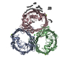
| Title | Cryo-EM structure of E. coli 70S ribosome bound to additional non-ribosomal proteins. | |||||||||
 Map data Map data | Cryo-EM structure of E. coli 70S ribosome bound to additional non-ribosomal proteins. MapI | |||||||||
 Sample Sample |
| |||||||||
 Keywords Keywords | ribosome / ribosome assembly / moonlighting proteins. | |||||||||
| Biological species |  | |||||||||
| Method | single particle reconstruction / cryo EM / Resolution: 12.9 Å | |||||||||
 Authors Authors | Shasmal M / Dey S / Shaikh TR / Bhakta S / Sengupta J | |||||||||
 Citation Citation |  Journal: Sci Rep / Year: 2016 Journal: Sci Rep / Year: 2016Title: E. coli metabolic protein aldehyde-alcohol dehydrogenase-E binds to the ribosome: a unique moonlighting action revealed. Authors: Manidip Shasmal / Sandip Dey / Tanvir R Shaikh / Sayan Bhakta / Jayati Sengupta /   Abstract: It is becoming increasingly evident that a high degree of regulation is involved in the protein synthesis machinery entailing more interacting regulatory factors. A multitude of proteins have been ...It is becoming increasingly evident that a high degree of regulation is involved in the protein synthesis machinery entailing more interacting regulatory factors. A multitude of proteins have been identified recently which show regulatory function upon binding to the ribosome. Here, we identify tight association of a metabolic protein aldehyde-alcohol dehydrogenase E (AdhE) with the E. coli 70S ribosome isolated from cell extract under low salt wash conditions. Cryo-EM reconstruction of the ribosome sample allows us to localize its position on the head of the small subunit, near the mRNA entrance. Our study demonstrates substantial RNA unwinding activity of AdhE which can account for the ability of ribosome to translate through downstream of at least certain mRNA helices. Thus far, in E. coli, no ribosome-associated factor has been identified that shows downstream mRNA helicase activity. Additionally, the cryo-EM map reveals interaction of another extracellular protein, outer membrane protein C (OmpC), with the ribosome at the peripheral solvent side of the 50S subunit. Our result also provides important insight into plausible functional role of OmpC upon ribosome binding. Visualization of the ribosome purified directly from the cell lysate unveils for the first time interactions of additional regulatory proteins with the ribosome. | |||||||||
| History |
|
- Structure visualization
| Movie |
 Movie viewer Movie viewer |
|---|---|
| Structure viewer | EM map:  SurfView SurfView Molmil Molmil Jmol/JSmol Jmol/JSmol |
| Supplemental images |
- Downloads & links
-EMDB archive
| Map data |  emd_2970.map.gz emd_2970.map.gz | 43.3 MB |  EMDB map data format EMDB map data format | |
|---|---|---|---|---|
| Header (meta data) |  emd-2970-v30.xml emd-2970-v30.xml emd-2970.xml emd-2970.xml | 9.6 KB 9.6 KB | Display Display |  EMDB header EMDB header |
| Images |  Image_MapI_EMD2970.tif Image_MapI_EMD2970.tif | 150 KB | ||
| Archive directory |  http://ftp.pdbj.org/pub/emdb/structures/EMD-2970 http://ftp.pdbj.org/pub/emdb/structures/EMD-2970 ftp://ftp.pdbj.org/pub/emdb/structures/EMD-2970 ftp://ftp.pdbj.org/pub/emdb/structures/EMD-2970 | HTTPS FTP |
-Related structure data
- Links
Links
| EMDB pages |  EMDB (EBI/PDBe) / EMDB (EBI/PDBe) /  EMDataResource EMDataResource |
|---|---|
| Related items in Molecule of the Month |
- Map
Map
| File |  Download / File: emd_2970.map.gz / Format: CCP4 / Size: 45.3 MB / Type: IMAGE STORED AS FLOATING POINT NUMBER (4 BYTES) Download / File: emd_2970.map.gz / Format: CCP4 / Size: 45.3 MB / Type: IMAGE STORED AS FLOATING POINT NUMBER (4 BYTES) | ||||||||||||||||||||||||||||||||||||||||||||||||||||||||||||
|---|---|---|---|---|---|---|---|---|---|---|---|---|---|---|---|---|---|---|---|---|---|---|---|---|---|---|---|---|---|---|---|---|---|---|---|---|---|---|---|---|---|---|---|---|---|---|---|---|---|---|---|---|---|---|---|---|---|---|---|---|---|
| Annotation | Cryo-EM structure of E. coli 70S ribosome bound to additional non-ribosomal proteins. MapI | ||||||||||||||||||||||||||||||||||||||||||||||||||||||||||||
| Projections & slices | Image control
Images are generated by Spider. | ||||||||||||||||||||||||||||||||||||||||||||||||||||||||||||
| Voxel size | X=Y=Z: 1.69 Å | ||||||||||||||||||||||||||||||||||||||||||||||||||||||||||||
| Density |
| ||||||||||||||||||||||||||||||||||||||||||||||||||||||||||||
| Symmetry | Space group: 1 | ||||||||||||||||||||||||||||||||||||||||||||||||||||||||||||
| Details | EMDB XML:
CCP4 map header:
| ||||||||||||||||||||||||||||||||||||||||||||||||||||||||||||
-Supplemental data
- Sample components
-Entire : E. coli 70S ribosome
| Entire | Name: E. coli 70S ribosome |
|---|---|
| Components |
|
-Supramolecule #1000: E. coli 70S ribosome
| Supramolecule | Name: E. coli 70S ribosome / type: sample / ID: 1000 / Details: Monodisperse, single particle form / Number unique components: 1 |
|---|---|
| Molecular weight | Theoretical: 2.5 MDa |
-Supramolecule #1: Escherichia coli 70S ribosome
| Supramolecule | Name: Escherichia coli 70S ribosome / type: complex / ID: 1 / Name.synonym: 70S ribosome / Recombinant expression: No / Ribosome-details: ribosome-prokaryote: ALL |
|---|---|
| Source (natural) | Organism:  |
| Molecular weight | Theoretical: 2.5 MDa |
-Experimental details
-Structure determination
| Method | cryo EM |
|---|---|
 Processing Processing | single particle reconstruction |
| Aggregation state | particle |
- Sample preparation
| Buffer | pH: 7.5 Details: 20 mM Tris-HCl, pH 7.5, 10 mM MgOAc, 30 mM NH4Cl, 5 mM 2-mercaptoethanol |
|---|---|
| Grid | Details: 300 mesh carbon coated lacey copper grid |
| Vitrification | Cryogen name: ETHANE / Chamber humidity: 100 % / Chamber temperature: 90 K / Instrument: GATAN CRYOPLUNGE 3 / Method: Blot for 4.3 seconds before plunging |
- Electron microscopy
| Microscope | FEI TECNAI F20 |
|---|---|
| Temperature | Min: 85 K / Max: 95 K |
| Alignment procedure | Legacy - Astigmatism: Astigmatism was corrected at 90000 magnification. |
| Details | Low dose imaging |
| Date | Mar 21, 2013 |
| Image recording | Category: CCD / Film or detector model: TVIPS TEMCAM-F415 (4k x 4k) / Digitization - Sampling interval: 4 µm / Number real images: 271 / Average electron dose: 15 e/Å2 |
| Electron beam | Acceleration voltage: 200 kV / Electron source:  FIELD EMISSION GUN FIELD EMISSION GUN |
| Electron optics | Calibrated magnification: 88466 / Illumination mode: FLOOD BEAM / Imaging mode: BRIGHT FIELD / Cs: 1.2 mm / Nominal defocus max: 4.6 µm / Nominal defocus min: 1.5 µm / Nominal magnification: 89000 |
| Sample stage | Specimen holder model: GATAN LIQUID NITROGEN |
| Experimental equipment |  Model: Tecnai F20 / Image courtesy: FEI Company |
- Image processing
| Details | Particles are selected first automatically by SPIDER and then manually by Jweb, grouped on the basis of defocus values and aligned by projection matching |
|---|---|
| CTF correction | Details: CTF correction of 3D map |
| Final reconstruction | Applied symmetry - Point group: C1 (asymmetric) / Algorithm: OTHER / Resolution.type: BY AUTHOR / Resolution: 12.9 Å / Resolution method: OTHER / Software - Name: SPIDER / Number images used: 56165 |
-Atomic model buiding 1
| Initial model | PDB ID: Chain - Chain ID: A |
|---|---|
| Software | Name: PyMOL, MDFF |
| Details | First manual rigid body fitting followed by flexible fitting by MDFF. |
| Refinement | Space: REAL / Protocol: FLEXIBLE FIT |
 Movie
Movie Controller


 Z (Sec.)
 Y (Row.)
 X (Col.)


