Movie
Movie Controller

[English] 日本語
 Yorodumi
- EMDB-1564: Recognition of Phe-tRNAPhe, Trp-tRNATrp and : a common molecular ... -
+ Open data

- Basic information
| Entry | Database: EMDB / ID: EMD-1564 | |||||||||
|---|---|---|---|---|---|---|---|---|---|---|
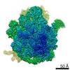
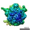
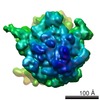
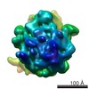
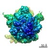
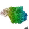
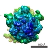
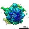
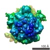
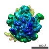
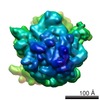
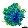
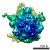
| Title | Recognition of Phe-tRNAPhe, Trp-tRNATrp and : a common molecular mechanism for aminoacyl-tRNA selection revealed by cryo-EM | |||||||||
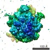
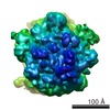
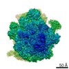
 Map data Map data | Leu-tRNA-EFTu-kir-GDP-70S ribosome | |||||||||
 Sample Sample |
| |||||||||
 Keywords Keywords | protein translation / decoding / elongation factor Tu / ribosome | |||||||||
| Function / homology |  Function and homology information Function and homology informationguanyl-nucleotide exchange factor complex / protein-synthesizing GTPase / guanosine tetraphosphate binding / stringent response / translational elongation / misfolded RNA binding / Group I intron splicing / RNA folding / translation elongation factor activity / translational termination ...guanyl-nucleotide exchange factor complex / protein-synthesizing GTPase / guanosine tetraphosphate binding / stringent response / translational elongation / misfolded RNA binding / Group I intron splicing / RNA folding / translation elongation factor activity / translational termination / positive regulation of RNA splicing / maintenance of translational fidelity / ribosomal large subunit assembly / large ribosomal subunit rRNA binding / cytosolic small ribosomal subunit / cytosolic large ribosomal subunit / cytoplasmic translation / tRNA binding / rRNA binding / structural constituent of ribosome / ribosome / translation / response to antibiotic / GTPase activity / GTP binding / magnesium ion binding / RNA binding / plasma membrane / cytoplasm / cytosol Similarity search - Function | |||||||||
| Biological species |  | |||||||||
| Method | single particle reconstruction / cryo EM / negative staining / Resolution: 12.0 Å | |||||||||
 Authors Authors | Li W / Agirrezabala X / Lei J / Bouakaz L / Brunelle JL / Ortiz-Meoz RF / Green R / Sanyal S / Ehrenberg M / Frank J | |||||||||
 Citation Citation | Journal: EMBO J / Year: 2008 Title: Recognition of aminoacyl-tRNA: a common molecular mechanism revealed by cryo-EM. Authors: Wen Li / Xabier Agirrezabala / Jianlin Lei / Lamine Bouakaz / Julie L Brunelle / Rodrigo F Ortiz-Meoz / Rachel Green / Suparna Sanyal / Måns Ehrenberg / Joachim Frank /  Abstract: The accuracy of ribosomal translation is achieved by an initial selection and a proofreading step, mediated by EF-Tu, which forms a ternary complex with aminoacyl(aa)-tRNA. To study the binding modes ...The accuracy of ribosomal translation is achieved by an initial selection and a proofreading step, mediated by EF-Tu, which forms a ternary complex with aminoacyl(aa)-tRNA. To study the binding modes of different aa-tRNAs, we compared cryo-EM maps of the kirromycin-stalled ribosome bound with ternary complexes containing Phe-tRNA(Phe), Trp-tRNA(Trp), or Leu-tRNA(LeuI). The three maps suggest a common binding manner of cognate aa-tRNAs in their specific binding with both the ribosome and EF-Tu. All three aa-tRNAs have the same 'loaded spring' conformation with a kink and twist between the D-stem and anticodon stem. The three complexes are similarly integrated in an interaction network, extending from the anticodon loop through h44 and protein S12 to the EF-Tu-binding CCA end of aa-tRNA, proposed to signal cognate codon-anticodon interaction to the GTPase centre and tune the accuracy of aa-tRNA selection. | |||||||||
| History |
|
- Structure visualization
| Movie |
Movie viewer |
|---|---|
| Structure viewer | EM map: SurfViewMolmilJmol/JSmol |
| Supplemental images |

- Downloads & links
-EMDB archive
| Map data | emd_1564.map.gz | 7.7 MB | EMDB map data format | |
|---|---|---|---|---|
| Header (meta data) | emd-1564-v30.xmlemd-1564.xml | 11.4 KB 11.4 KB | Display Display | EMDB header |
| Images | 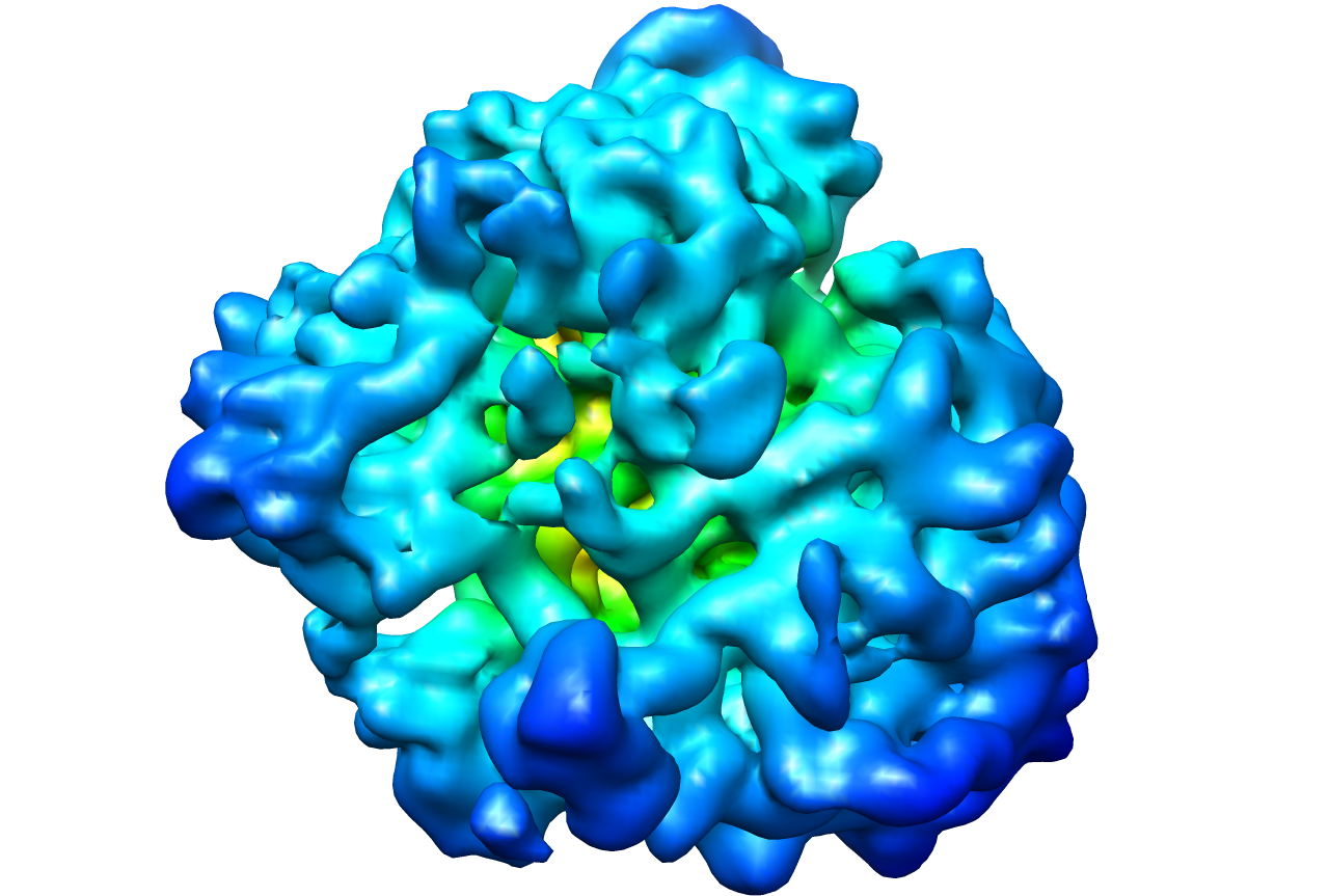 1564.png 1564.png | 551.1 KB | ||
| Archive directory |  http://ftp.pdbj.org/pub/emdb/structures/EMD-1564ftp://ftp.pdbj.org/pub/emdb/structures/EMD-1564 http://ftp.pdbj.org/pub/emdb/structures/EMD-1564ftp://ftp.pdbj.org/pub/emdb/structures/EMD-1564 | HTTPS FTP |
-Related structure data
| Related structure data |  3eq4MC 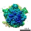 1565C 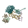 3ep2C 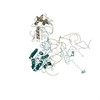 3eq3C M: atomic model generated by this map C: citing same article ( |
|---|---|
| Similar structure data |

-Links
| EMDB pages | EMDB (EBI/PDBe) / EMDataResource |
|---|---|
| Related items in Molecule of the Month |


-Map
| File | Download / File: emd_1564.map.gz / Format: CCP4 / Size: 8.2 MB / Type: IMAGE STORED AS FLOATING POINT NUMBER (4 BYTES) | ||||||||||||||||||||||||||||||||||||||||||||||||||||||||||||||||||||
|---|---|---|---|---|---|---|---|---|---|---|---|---|---|---|---|---|---|---|---|---|---|---|---|---|---|---|---|---|---|---|---|---|---|---|---|---|---|---|---|---|---|---|---|---|---|---|---|---|---|---|---|---|---|---|---|---|---|---|---|---|---|---|---|---|---|---|---|---|---|
| Annotation | Leu-tRNA-EFTu-kir-GDP-70S ribosome | ||||||||||||||||||||||||||||||||||||||||||||||||||||||||||||||||||||
| Projections & slices | Image control
Images are generated by Spider. | ||||||||||||||||||||||||||||||||||||||||||||||||||||||||||||||||||||
| Voxel size | X=Y=Z: 2.82 Å | ||||||||||||||||||||||||||||||||||||||||||||||||||||||||||||||||||||
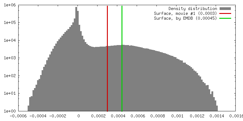
| Density |
| ||||||||||||||||||||||||||||||||||||||||||||||||||||||||||||||||||||
| Symmetry | Space group: 1 | ||||||||||||||||||||||||||||||||||||||||||||||||||||||||||||||||||||
| Details | EMDB XML:
CCP4 map header:
| ||||||||||||||||||||||||||||||||||||||||||||||||||||||||||||||||||||
 Z (Sec.)
 Y (Row.)
 X (Col.)


-Supplemental data
- Sample components
-Entire : Leu-tRNA-EFTu-GDP-kir-70S ribosome
| Entire | Name: Leu-tRNA-EFTu-GDP-kir-70S ribosome |
|---|---|
| Components |
|
-Supramolecule #1000: Leu-tRNA-EFTu-GDP-kir-70S ribosome
| Supramolecule | Name: Leu-tRNA-EFTu-GDP-kir-70S ribosome / type: sample / ID: 1000 / Details: sedimentation / Oligomeric state: monomeric / Number unique components: 5 |
|---|---|
| Molecular weight | Experimental: 2.8 MDa / Theoretical: 2.8 MDa |
-Supramolecule #1: 70S ribosome
| Supramolecule | Name: 70S ribosome / type: complex / ID: 1 / Name.synonym: 70S ribosome / Recombinant expression: No / Ribosome-details: ribosome-prokaryote: ALL |
|---|---|
| Source (natural) | Organism: |
| Molecular weight | Experimental: 2.8 MDa / Theoretical: 2.8 MDa |
-Experimental details
-Structure determination
| Method | negative staining, cryo EM |
|---|---|
 Processing Processing | single particle reconstruction |
| Aggregation state | particle |
-Sample preparation
| Concentration | 0.03 mg/mL |
|---|---|
| Buffer | pH: 7.3 Details: Polymix buffer( 5 mM potassium phosphate (KH2PO4) pH 7.3, 5 mM NH4Cl, 95 mM KCl, 0.5 mM CaCl2, 8 mM putrescine, 1 mM spermidine, 1 mM DTE, 5 mM magnesium acetate) |
| Staining | Type: NEGATIVE / Details: cryo |
| Grid | Details: 200 mesh |
| Vitrification | Cryogen name: NITROGEN / Chamber humidity: 90 % / Chamber temperature: 80 K / Instrument: OTHER / Details: Vitrification instrument: vitrobot / Method: Blot for 6 seconds |
- Electron microscopy
| Microscope | FEI TECNAI F20 |
|---|---|
| Temperature | Min: 80.7 K / Max: 80.7 K / Average: 80.7 K |
| Alignment procedure | Legacy - Astigmatism: corrected objective at 100,000 times magnification Legacy - Electron beam tilt params: no tilt |
| Specialist optics | Energy filter - Name: no energy filter |
| Date | Jun 1, 2006 |
| Image recording | Category: FILM / Film or detector model: KODAK SO-163 FILM / Digitization - Scanner: ZEISS SCAI / Digitization - Sampling interval: 7 µm / Number real images: 112 / Average electron dose: 15 e/Å2 / Bits/pixel: 2 |
| Tilt angle min | 0 |
| Tilt angle max | 0 |
| Electron beam | Acceleration voltage: 300 kV / Electron source:  FIELD EMISSION GUN FIELD EMISSION GUN |
| Electron optics | Calibrated magnification: 49645 / Illumination mode: FLOOD BEAM / Imaging mode: BRIGHT FIELD / Cs: 2.26 mm / Nominal defocus max: 4.0 µm / Nominal defocus min: 1.0 µm / Nominal magnification: 50000 |
| Sample stage | Specimen holder: cartridge / Specimen holder model: OTHER |
| Experimental equipment |  Model: Tecnai F20 / Image courtesy: FEI Company |
-Image processing
| CTF correction | Details: defocus groups, Wiener filter |
|---|---|
| Final reconstruction | Applied symmetry - Point group: C1 (asymmetric) / Algorithm: OTHER / Resolution.type: BY AUTHOR / Resolution: 12.0 Å / Resolution method: FSC 0.5 CUT-OFF / Software - Name: Spider / Number images used: 80000 |
| Final angle assignment | Details: spider |
-Atomic model buiding 1
| Initial model | (PDB ID:  2avy 2aw4 1gix , ) |
|---|---|
| Software | Name: TNT |
| Details | Protocol: real space refinement. 1lBM(1989-1996 fragment) |
| Refinement | Space: REAL / Protocol: RIGID BODY FIT |
| Output model | PDB-3eq4: |