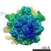
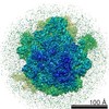
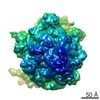
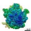
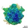
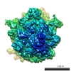
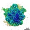
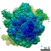
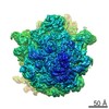
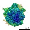
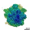
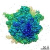
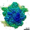
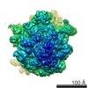
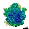
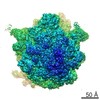
- EMDB-1003: Solution structure of the E. coli 70S ribosome at 11.5 A resolution. -
+
Open data
ID or keywords:
Loading...
-
Basic information
Entry
Database: EMDB / ID: EMD-1003
Title
Solution structure of the E. coli 70S ribosome at 11.5 A resolution.
Map data
E. coli 70S Ribosome
Sample
Sample: FMet-tRNAMet 70S Ribosome from E.coli
Complex: 70S ribosome Escherichia coli
RNA: fMet-tRNA
RNA: MF-mRNA
Function / homology
Function and homology information
regulation of translation / large ribosomal subunit / ribosome biogenesis / ribosomal small subunit biogenesis / small ribosomal subunit / small ribosomal subunit rRNA binding / large ribosomal subunit rRNA binding / cytosolic small ribosomal subunit / cytosolic large ribosomal subunit / cytoplasmic translation ...regulation of translation / large ribosomal subunit / ribosome biogenesis / ribosomal small subunit biogenesis / small ribosomal subunit / small ribosomal subunit rRNA binding / large ribosomal subunit rRNA binding / cytosolic small ribosomal subunit / cytosolic large ribosomal subunit / cytoplasmic translation / tRNA binding / rRNA binding / structural constituent of ribosome / ribosome / translation / ribonucleoprotein complex / response to antibiotic / cytoplasm Similarity search - Function
Ribosomal protein L1, bacterial-type / Ribosomal protein L1, conserved site / Ribosomal protein L1 signature. / Ribosomal protein L1 / Ribosomal protein L1, 3-layer alpha/beta-sandwich / Ribosomal protein L1-like / Ribosomal protein L1/ribosomal biogenesis protein / Ribosomal protein L1p/L10e family / Ribosomal protein L11, bacterial-type / Ribosomal protein L11, conserved site ...Ribosomal protein L1, bacterial-type / Ribosomal protein L1, conserved site / Ribosomal protein L1 signature. / Ribosomal protein L1 / Ribosomal protein L1, 3-layer alpha/beta-sandwich / Ribosomal protein L1-like / Ribosomal protein L1/ribosomal biogenesis protein / Ribosomal protein L1p/L10e family / Ribosomal protein L11, bacterial-type / Ribosomal protein L11, conserved site / Ribosomal protein L11 signature. / Ribosomal protein L6, conserved site / Ribosomal protein L6 signature 1. / Ribosomal protein L11, N-terminal / Ribosomal protein L11, N-terminal domain / Ribosomal protein L11/L12 / Ribosomal protein L11, C-terminal / Ribosomal protein L11, C-terminal domain superfamily / Ribosomal protein L11/L12, N-terminal domain superfamily / Ribosomal protein L11/L12 / Ribosomal protein L11, RNA binding domain / Ribosomal protein S6, conserved site / Ribosomal protein S6 signature. / Ribosomal protein L6, bacterial-type / Ribosomal protein S7, bacterial/organellar-type / Ribosomal protein S4, bacterial-type / Ribosomal protein S5, bacterial-type / 30S ribosomal protein S17 / Ribosomal protein S6, plastid/chloroplast / Ribosomal protein S15, bacterial-type / Ribosomal protein S6 / Ribosomal protein S6 / Ribosomal protein S6 superfamily / Translation elongation factor EF1B/ribosomal protein S6 / Ribosomal protein S5, N-terminal, conserved site / Ribosomal protein S5 signature. / Ribosomal protein S7, conserved site / Ribosomal protein S7 signature. / : / Ribosomal protein L6, alpha-beta domain / Ribosomal protein L6 / Ribosomal protein L6 / Ribosomal protein L6, alpha-beta domain superfamily / Ribosomal protein S17, conserved site / Ribosomal protein S17 signature. / Ribosomal protein S5 / S5 double stranded RNA-binding domain profile. / Ribosomal protein S5, N-terminal / Ribosomal protein S5, C-terminal / Ribosomal protein S5, N-terminal domain / Ribosomal protein S5, C-terminal domain / Ribosomal protein S8 signature. / Ribosomal protein S4/S9 N-terminal domain / Ribosomal protein S4, conserved site / Ribosomal protein S4 signature. / Ribosomal protein S4/S9 N-terminal domain / Ribosomal protein S4/S9, N-terminal / Ribosomal protein S15 signature. / Ribosomal protein S4/S9 / Ribosomal protein S8 / Ribosomal protein S8 superfamily / Ribosomal protein S8 / S4 RNA-binding domain profile. / S4 RNA-binding domain / S4 domain / RNA-binding S4 domain / RNA-binding S4 domain superfamily / Ribosomal protein S5/S7 / Ribosomal protein S7 domain / Ribosomal protein S7 domain superfamily / Ribosomal protein S7p/S5e / Ribosomal protein S17/S11 / Ribosomal protein S17 / Ribosomal protein S15 / Ribosomal_S15 / Ribosomal protein S15 / S15/NS1, RNA-binding / Ribosomal protein S5 domain 2-type fold, subgroup / Ribosomal protein S5 domain 2-type fold / Nucleic acid-binding, OB-fold Similarity search - Domain/homology
Small ribosomal subunit protein uS5 / Large ribosomal subunit protein uL6 / Small ribosomal subunit protein uS8 / Small ribosomal subunit protein uS7 / Small ribosomal subunit protein uS4 / Small ribosomal subunit protein uS5 / Small ribosomal subunit protein bS6 / Small ribosomal subunit protein uS17 / Large ribosomal subunit protein uL1 / Large ribosomal subunit protein uL11 ...Small ribosomal subunit protein uS5 / Large ribosomal subunit protein uL6 / Small ribosomal subunit protein uS8 / Small ribosomal subunit protein uS7 / Small ribosomal subunit protein uS4 / Small ribosomal subunit protein uS5 / Small ribosomal subunit protein bS6 / Small ribosomal subunit protein uS17 / Large ribosomal subunit protein uL1 / Large ribosomal subunit protein uL11 / Small ribosomal subunit protein uS15 / Small ribosomal subunit protein uS4 / Large ribosomal subunit protein uL6 / Small ribosomal subunit protein uS15 / Large ribosomal subunit protein uL1 / Small ribosomal subunit protein bS6 Similarity search - Component
Biological species
Escherichia coli (E. coli)
Method
single particle reconstruction / cryo EM / Resolution: 11.5 Å
Journal: Cell / Year: 2000 Title: Solution structure of the E. coli 70S ribosome at 11.5 A resolution. Authors: I S Gabashvili / R K Agrawal / C M Spahn / R A Grassucci / D I Svergun / J Frank / P Penczek / Abstract: Over 73,000 projections of the E. coli ribosome bound with formyl-methionyl initiator tRNAf(Met) were used to obtain an 11.5 A cryo-electron microscopy map of the complex. This map allows ...Over 73,000 projections of the E. coli ribosome bound with formyl-methionyl initiator tRNAf(Met) were used to obtain an 11.5 A cryo-electron microscopy map of the complex. This map allows identification of RNA helices, peripheral proteins, and intersubunit bridges. Comparison of double-stranded RNA regions and positions of proteins identified in both cryo-EM and X-ray maps indicates good overall agreement but points to rearrangements of ribosomal components required for the subunit association. Fitting of known components of the 50S stalk base region into the map defines the architecture of the GTPase-associated center and reveals a major change in the orientation of the alpha-sarcin-ricin loop. Analysis of the bridging connections between the subunits provides insight into the dynamic signaling mechanism between the ribosomal subunits.
History
Deposition
Jun 26, 2002
-
Header (metadata) release
Aug 22, 2002
-
Map release
Aug 22, 2002
-
Update
Oct 17, 2012
-
Current status
Oct 17, 2012
Processing site: PDBe / Status: Released
-
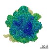
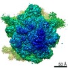
Structure visualization
Movie
Surface view with section colored by density value
Supramolecule #1000: FMet-tRNAMet 70S Ribosome from E.coli
Supramolecule
Name: FMet-tRNAMet 70S Ribosome from E.coli / type: sample / ID: 1000 Details: preparation and buffer conditions are as described in Malhotra et al., J. Mol. Biol. (1998) 280, 103-116 Number unique components: 3
Name: fMet-tRNA / type: rna / ID: 1 / Classification: OTHER / Structure: DOUBLE HELIX / Synthetic?: No
Source (natural)
Organism: Escherichia coli (E. coli)
-
Macromolecule #2: MF-mRNA
Macromolecule
Name: MF-mRNA / type: rna / ID: 2 / Details: 46 nucleotides / Classification: OTHER / Structure: SINGLE STRANDED / Synthetic?: No
Source (natural)
Organism: Escherichia coli (E. coli)
-
Experimental details
-
Structure determination
Method
cryo EM
Processing
single particle reconstruction
Aggregation state
particle
-
Sample preparation
Concentration
0.09 mg/mL
Buffer
pH: 7.6 Details: 20 mM Hepes, 6 mM Mg(CH3COO)2 150 mM NH4Cl, 4 mM beta-Mercaptoethanol, 0.5 mM Spermine, 2 mM Spermidine.
Vitrification
Cryogen name: ETHANE / Instrument: HOMEMADE PLUNGER / Details: Vitrification instrument: 2 side blotting plunger / Method: Blot and plunge
-
Electron microscopy
Microscope
FEI/PHILIPS CM200FEG/ST
Temperature
Average: 93 K
Details
25 % of data from Philips EM420 with LaB6 at 100kV
Date
Oct 4, 1997
Image recording
Category: FILM / Film or detector model: KODAK SO-163 FILM / Digitization - Scanner: PERKIN ELMER / Digitization - Sampling interval: 15 µm / Number real images: 239 / Average electron dose: 10 e/Å2 / Bits/pixel: 12
Tilt angle min
0
Tilt angle max
0
Electron beam
Acceleration voltage: 200 kV / Electron source: FIELD EMISSION GUN
Details: Wiener filtration of defocus group volumes
Final reconstruction
Applied symmetry - Point group: C1 (asymmetric) / Algorithm: OTHER / Resolution.type: BY AUTHOR / Resolution: 11.5 Å / Resolution method: FSC 0.5 CUT-OFF / Software - Name: SPIDER/WEB / Number images used: 73523
-
Atomic model buiding 1
Software
Name: O
Details
manual fitting using O Mol_Id: 1; Molecule: S4 Ribosomal Protein; Chain: A; Other_Details: Modeled By Analogous Protein Of B. Stearothermophilus Taken From Pdb Entry 1C06 Mol_Id: 2; Molecule: S5 Ribosomal Protein; Chain: B; Other_Details: Modeled By Analogous Protein Of B. Stearothermophilus Taken From Pdb Entry 1Pkp Mol_Id: 3; Molecule: S6 Ribosomal Protein; Chain: C; Other_Details: Modeled By Analogous Protein Of T. Thermophilus Taken From Pdb Entry 1Ris Mol_Id: 4; Molecule: S7 Ribosomal Protein; Chain: D; Other_Details: Modeled By Analogous Protein Of T. Thermophilus Taken From Pdb Entry 1Rss Mol_Id: 5; Molecule: S8 Ribosomal Protein; Chain: E; Other_Details: Modeled By Analogous Protein Of T. Thermophilus Taken From Pdb Entry 1An7 Mol_Id: 6; Molecule: S15 Ribosomal Protein; Chain: F; Other_Details: Modeled By Analogous Protein Of B. Stearothermophilus Taken From Pdb Entry 1A32 Mol_Id: 7; Molecule: S17 Ribosomal Protein; Chain: G; Other_Details: Modeled By Analogous Protein Of B. Stearothermophilus Taken From Pdb Entries 1Rip and 1Qd7 Mol_Id: 8; Molecule: S20 Ribosomal Protein; Chain: H; Other_Details: Modeled By Analogous Protein Of T. Thermophilus Taken From Pdb Entry 1Qd7 Mol_Id: 9; Molecule: Ribosomal Protein L1; Chain: N; Other_Details: Modeled By Analogous Protein Of T. Thermophilus Taken From Pdb Entry 1Ad2 Mol_Id: 10; Molecule: Ribosomal Protein L6; Chain: J; Mutation: Yes; Other_Details: Modeled By Analogous Protein Of T. Stearothermophilus Taken From Pdb Entry 1Rl6 Mol_Id: 11; Molecule: Ribosomal Protein L11; Chain: K; Other_Details: Modeled By Analogous Protein Of T. Maritima Taken From Pdb Entry 1Mms Mol_Id: 12; Molecule: Fragment Of 16S Rrna Helix 23; Chain: I; Fragment: Residues 673-713; Other_Details: Modeled As Analogous Fragment Of T. Thermophilus Taken From Pdb Entry 1Qd7 Mol_Id: 13; Molecule: Fragment Of 23S Rrna; Chain: L; Fragment: Residues 1051-1108; Other_Details: T. Maritima RNA Sequence and Model Taken From Pdb Entry 1Mms Mol_Id: 14; Molecule: Helix 95 Of 23S Rrna; Chain: M; Other_Details: E. Coli RNA Sequence and Model Taken From Pdb Entry 480D Mol_Id: 15; Molecule: Formyl-Methionyl-tRNA; Chain: O; Synonym: Fmet-tRNA; Other_Details: E. Coli Fmet-tRNA Sequence and Model Taken From Pdb Entry 2Fmt
Refinement
Protocol: RIGID BODY FIT
Output model
PDB-1eg0: FITTING OF COMPONENTS WITH KNOWN STRUCTURE INTO AN 11.5 A CRYO-EM MAP OF THE E.COLI 70S RIBOSOME
+
About Yorodumi
-
News
-
Feb 9, 2022. New format data for meta-information of EMDB entries
New format data for meta-information of EMDB entries
Version 3 of the EMDB header file is now the official format.
The previous official version 1.9 will be removed from the archive.
In the structure databanks used in Yorodumi, some data are registered as the other names, "COVID-19 virus" and "2019-nCoV". Here are the details of the virus and the list of structure data.
Jan 31, 2019. EMDB accession codes are about to change! (news from PDBe EMDB page)
EMDB accession codes are about to change! (news from PDBe EMDB page)
The allocation of 4 digits for EMDB accession codes will soon come to an end. Whilst these codes will remain in use, new EMDB accession codes will include an additional digit and will expand incrementally as the available range of codes is exhausted. The current 4-digit format prefixed with “EMD-” (i.e. EMD-XXXX) will advance to a 5-digit format (i.e. EMD-XXXXX), and so on. It is currently estimated that the 4-digit codes will be depleted around Spring 2019, at which point the 5-digit format will come into force.
The EM Navigator/Yorodumi systems omit the EMD- prefix.
Related info.:Q: What is EMD? / ID/Accession-code notation in Yorodumi/EM Navigator
Yorodumi is a browser for structure data from EMDB, PDB, SASBDB, etc.
This page is also the successor to EM Navigator detail page, and also detail information page/front-end page for Omokage search.
The word "yorodu" (or yorozu) is an old Japanese word meaning "ten thousand". "mi" (miru) is to see.
Related info.:EMDB / PDB / SASBDB / Comparison of 3 databanks / Yorodumi Search / Aug 31, 2016. New EM Navigator & Yorodumi / Yorodumi Papers / Jmol/JSmol / Function and homology information / Changes in new EM Navigator and Yorodumi
 Movie
Movie Controller

 Yorodumi
 Open data
 Basic information
 Map data
 Sample
 Function and homology information
 Authors
 Citation
 Structure visualization
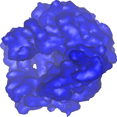
 Downloads & links
 1003.gif
 http://ftp.pdbj.org/pub/emdb/structures/EMD-1003


 Z (Sec.)
 Y (Row.)
 X (Col.)


 Sample components
 Processing
 Electron microscopy
 FIELD EMISSION GUN