Movie
Movie Controller

[English] 日本語
 Yorodumi
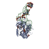
- EMDB-5036: Aminoacyl-tRNA-EF-Tu-GDP-kir ternary complex-bound E. coli 70S ri... -
+ Open data

- Basic information
| Entry | Database: EMDB / ID: EMD-5036 | |||||||||
|---|---|---|---|---|---|---|---|---|---|---|
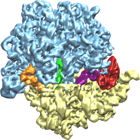
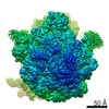
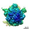
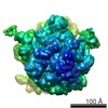
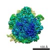
| Title | Aminoacyl-tRNA-EF-Tu-GDP-kir ternary complex-bound E. coli 70S ribosome | |||||||||
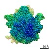
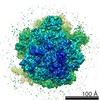
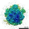
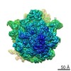
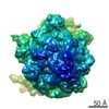
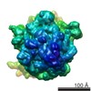
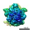
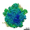
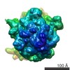
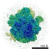
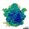
 Map data Map data | Ternary complex-bound 70S E. coli ribosome | |||||||||
 Sample Sample |
| |||||||||
 Keywords Keywords | decoding / tRNA selection / GTPase / accommodation / flexible fitting / cryo-EM / MDFF / hydrophobic gate / EF-Tu / ribosome / ternary complex | |||||||||
| Function / homology |  Function and homology information Function and homology informationguanyl-nucleotide exchange factor complex / protein-synthesizing GTPase / guanosine tetraphosphate binding / stringent response / transcription antitermination factor activity, RNA binding / ornithine decarboxylase inhibitor activity / translational elongation / misfolded RNA binding / Group I intron splicing / RNA folding ...guanyl-nucleotide exchange factor complex / protein-synthesizing GTPase / guanosine tetraphosphate binding / stringent response / transcription antitermination factor activity, RNA binding / ornithine decarboxylase inhibitor activity / translational elongation / misfolded RNA binding / Group I intron splicing / RNA folding / translation elongation factor activity / translational termination / transcriptional attenuation / endoribonuclease inhibitor activity / positive regulation of ribosome biogenesis / RNA-binding transcription regulator activity / four-way junction DNA binding / negative regulation of cytoplasmic translation / DnaA-L2 complex / regulation of mRNA stability / translation repressor activity / negative regulation of translational initiation / negative regulation of DNA-templated DNA replication initiation / mRNA regulatory element binding translation repressor activity / positive regulation of RNA splicing / regulation of DNA-templated transcription elongation / transcription elongation factor complex / response to reactive oxygen species / cytosolic ribosome assembly / ribosome assembly / assembly of large subunit precursor of preribosome / transcription antitermination / DNA endonuclease activity / regulation of cell growth / DNA-templated transcription termination / response to radiation / maintenance of translational fidelity / mRNA 5'-UTR binding / regulation of translation / large ribosomal subunit / transferase activity / ribosomal small subunit assembly / ribosome biogenesis / ribosome binding / ribosomal small subunit biogenesis / 5S rRNA binding / ribosomal large subunit assembly / small ribosomal subunit / small ribosomal subunit rRNA binding / large ribosomal subunit rRNA binding / cytosolic small ribosomal subunit / cytosolic large ribosomal subunit / cytoplasmic translation / tRNA binding / negative regulation of translation / rRNA binding / structural constituent of ribosome / ribosome / translation / response to antibiotic / negative regulation of DNA-templated transcription / hydrolase activity / mRNA binding / GTPase activity / GTP binding / magnesium ion binding / DNA binding / RNA binding / zinc ion binding / membrane / plasma membrane / cytoplasm / cytosol Similarity search - Function | |||||||||
| Biological species |  | |||||||||
| Method | single particle reconstruction / cryo EM / Resolution: 6.7 Å | |||||||||
 Authors Authors | Villa E / Sengupta J / Trabuco LG / LeBarron J / Baxter WT / Shaikh TR / Grassucci RA / Nissen P / Ehrenberg M / Schulten K / Frank J | |||||||||
 Citation Citation | Journal: Structure / Year: 2008 Title: Flexible fitting of atomic structures into electron microscopy maps using molecular dynamics. Authors: Leonardo G Trabuco / Elizabeth Villa / Kakoli Mitra / Joachim Frank / Klaus Schulten /  Abstract: A novel method to flexibly fit atomic structures into electron microscopy (EM) maps using molecular dynamics simulations is presented. The simulations incorporate the EM data as an external potential ...A novel method to flexibly fit atomic structures into electron microscopy (EM) maps using molecular dynamics simulations is presented. The simulations incorporate the EM data as an external potential added to the molecular dynamics force field, allowing all internal features present in the EM map to be used in the fitting process, while the model remains fully flexible and stereochemically correct. The molecular dynamics flexible fitting (MDFF) method is validated for available crystal structures of protein and RNA in different conformations; measures to assess and monitor the fitting process are introduced. The MDFF method is then used to obtain high-resolution structures of the E. coli ribosome in different functional states imaged by cryo-EM. | |||||||||
| History |
|
- Structure visualization
| Movie |
Movie viewer |
|---|---|
| Structure viewer | EM map: SurfViewMolmilJmol/JSmol |
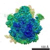
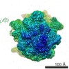
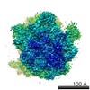
| Supplemental images |
- Downloads & links
-EMDB archive
| Map data | emd_5036.map.gz | 104.3 MB | EMDB map data format | |
|---|---|---|---|---|
| Header (meta data) | emd-5036-v30.xmlemd-5036.xml | 17.1 KB 17.1 KB | Display Display | EMDB header |
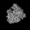
| Images |  emd_5036_1.png emd_5036_1.png | 275.3 KB | ||
| Archive directory |  http://ftp.pdbj.org/pub/emdb/structures/EMD-5036ftp://ftp.pdbj.org/pub/emdb/structures/EMD-5036 http://ftp.pdbj.org/pub/emdb/structures/EMD-5036ftp://ftp.pdbj.org/pub/emdb/structures/EMD-5036 | HTTPS FTP |
-Related structure data
| Related structure data |  4v69MC M: atomic model generated by this map C: citing same article ( |
|---|---|
| Similar structure data |
-Links
| EMDB pages | EMDB (EBI/PDBe) / EMDataResource |
|---|---|
| Related items in Molecule of the Month |


-Map
| File | Download / File: emd_5036.map.gz / Format: CCP4 / Size: 109.9 MB / Type: IMAGE STORED AS FLOATING POINT NUMBER (4 BYTES) | ||||||||||||||||||||||||||||||||||||||||||||||||||||||||||||||||||||
|---|---|---|---|---|---|---|---|---|---|---|---|---|---|---|---|---|---|---|---|---|---|---|---|---|---|---|---|---|---|---|---|---|---|---|---|---|---|---|---|---|---|---|---|---|---|---|---|---|---|---|---|---|---|---|---|---|---|---|---|---|---|---|---|---|---|---|---|---|---|
| Annotation | Ternary complex-bound 70S E. coli ribosome | ||||||||||||||||||||||||||||||||||||||||||||||||||||||||||||||||||||
| Projections & slices | Image control
Images are generated by Spider. | ||||||||||||||||||||||||||||||||||||||||||||||||||||||||||||||||||||
| Voxel size | X=Y=Z: 1.2 Å | ||||||||||||||||||||||||||||||||||||||||||||||||||||||||||||||||||||
| Density |
| ||||||||||||||||||||||||||||||||||||||||||||||||||||||||||||||||||||
| Symmetry | Space group: 1 | ||||||||||||||||||||||||||||||||||||||||||||||||||||||||||||||||||||
| Details | EMDB XML:
CCP4 map header:
| ||||||||||||||||||||||||||||||||||||||||||||||||||||||||||||||||||||
 Z (Sec.)
 Y (Row.)
 X (Col.)


-Supplemental data
- Sample components
-Entire : 70S ribosome from E. coli complex 70S-fMet-tRNA-Phe-tRNA-EF-Tu-GD...
| Entire | Name: 70S ribosome from E. coli complex 70S-fMet-tRNA-Phe-tRNA-EF-Tu-GDP-kirromycin. |
|---|---|
| Components |
|
-Supramolecule #1000: 70S ribosome from E. coli complex 70S-fMet-tRNA-Phe-tRNA-EF-Tu-GD...
| Supramolecule | Name: 70S ribosome from E. coli complex 70S-fMet-tRNA-Phe-tRNA-EF-Tu-GDP-kirromycin. type: sample / ID: 1000 / Oligomeric state: single particle / Number unique components: 5 |
|---|
-Supramolecule #1: 70S ribosome
| Supramolecule | Name: 70S ribosome / type: complex / ID: 1 / Recombinant expression: No / Database: NCBI / Ribosome-details: ribosome-prokaryote: ALL |
|---|---|
| Source (natural) | Organism: |
-Macromolecule #1: fMet-tRNAfMet
| Macromolecule | Name: fMet-tRNAfMet / type: rna / ID: 1 / Classification: TRANSFER / Structure: SINGLE STRANDED / Synthetic?: No |
|---|---|
| Source (natural) | Organism: |
-Macromolecule #2: Phe-tRNAPhe
| Macromolecule | Name: Phe-tRNAPhe / type: rna / ID: 2 / Classification: TRANSFER / Structure: SINGLE STRANDED / Synthetic?: No |
|---|---|
| Source (natural) | Organism: |
-Macromolecule #3: tRNA
| Macromolecule | Name: tRNA / type: rna / ID: 3 / Name.synonym: deacylated tRNA / Classification: TRANSFER / Structure: SINGLE STRANDED / Synthetic?: No |
|---|---|
| Source (natural) | Organism: |
-Macromolecule #4: Elongation factor Tu
| Macromolecule | Name: Elongation factor Tu / type: protein_or_peptide / ID: 4 / Name.synonym: EF-Tu / Details: EF-Tu bound to kirromycin and GDP / Number of copies: 1 / Oligomeric state: monomer / Recombinant expression: Yes |
|---|---|
| Source (natural) | Organism: |
| Recombinant expression | Organism: |
-Experimental details
-Structure determination
| Method | cryo EM |
|---|---|
 Processing Processing | single particle reconstruction |
| Aggregation state | particle |
-Sample preparation
| Concentration | 0.0768 mg/mL |
|---|---|
| Buffer | pH: 7.5 Details: 5 mM potassium phosphate, 5 mM magnesium acetate, 5 mM ammonium chloride, 95 mM potassium chloride, 0.5 mM calcium chloride, 8 mM putrescine, 1 mM spermidine, and 1 mM dithioerythritol |
| Grid | Details: Thin Carbon on 300 mesh Quantifoil R2/4 |
| Vitrification | Cryogen name: ETHANE / Chamber humidity: 90 % / Chamber temperature: 80 K / Instrument: OTHER Details: Vitrification instrument: FEI vitrobot. Blot Offset at -1 mm Method: Blot 3 seconds before plunging |
- Electron microscopy
| Microscope | FEI POLARA 300 |
|---|---|
| Temperature | Average: 84 K |
| Alignment procedure | Legacy - Astigmatism: Objective lens astigmatism was corrected at 115,000 times magnification with fastscan ccd |
| Details | Low dose |
| Date | Aug 31, 2006 |
| Image recording | Category: FILM / Film or detector model: KODAK SO-163 FILM / Digitization - Scanner: ZEISS SCAI / Digitization - Sampling interval: 7 µm / Number real images: 304 / Average electron dose: 20 e/Å2 / Od range: 1.2 / Bits/pixel: 12 |
| Electron beam | Acceleration voltage: 300 kV / Electron source:  FIELD EMISSION GUN FIELD EMISSION GUN |
| Electron optics | Calibrated magnification: 58279 / Illumination mode: FLOOD BEAM / Imaging mode: BRIGHT FIELD / Cs: 2.26 mm / Nominal defocus max: 4.52 µm / Nominal defocus min: 1.2 µm / Nominal magnification: 59000 |
| Sample stage | Specimen holder: FEI Polara Cartridge / Specimen holder model: OTHER |
| Experimental equipment |  Model: Tecnai Polara / Image courtesy: FEI Company |
-Image processing
| CTF correction | Details: Correction of reconstruction of each defocus group |
|---|---|
| Final reconstruction | Algorithm: OTHER / Resolution.type: BY AUTHOR / Resolution: 6.7 Å / Resolution method: OTHER / Software - Name: Spider / Number images used: 131599 |
-Atomic model buiding 1
| Initial model | PDB ID:  2i2u |
|---|---|
| Software | Name: MDFF |
| Details | Protocol: MDFF. An atomic model of the entire ribosome and factors was creating using molecular dynamics flexible fitting (Trabuco et al. Flexible Fitting of Atomic Structures into Electron Microscopy Maps Using Molecular Dynamics. Structure (2008) vol. 16 (5) pp. 673-683) |
| Refinement | Space: REAL / Protocol: FLEXIBLE FIT / Target criteria: RMSD, cross correlation |
| Output model | PDB-4v69: |
-Atomic model buiding 2
| Initial model | PDB ID: 2i2v |
|---|---|
| Software | Name: MDFF |
| Details | Protocol: MDFF. An atomic model of the entire ribosome and factors was creating using molecular dynamics flexible fitting (Trabuco et al. Flexible Fitting of Atomic Structures into Electron Microscopy Maps Using Molecular Dynamics. Structure (2008) vol. 16 (5) pp. 673-683) |
| Refinement | Space: REAL / Protocol: FLEXIBLE FIT / Target criteria: RMSD, cross correlation |
| Output model | PDB-4v69: |
-Atomic model buiding 3
| Initial model | PDB ID: |
|---|---|
| Software | Name: MDFF |
| Details | Protocol: MDFF. An atomic model of the entire ribosome and factors was creating using molecular dynamics flexible fitting (Trabuco et al. Flexible Fitting of Atomic Structures into Electron Microscopy Maps Using Molecular Dynamics. Structure (2008) vol. 16 (5) pp. 673-683) |
| Refinement | Space: REAL / Protocol: FLEXIBLE FIT / Target criteria: RMSD, cross correlation |
| Output model | PDB-4v69: |