 ムービー
ムービー コントローラー


+ データを開く

- 基本情報
基本情報
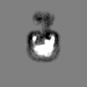
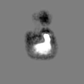
| 登録情報 | データベース: EMDB / ID: EMD-2927 | |||||||||
|---|---|---|---|---|---|---|---|---|---|---|
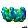
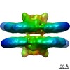
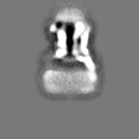
| タイトル | Negative stain structure of a type 6 secretion system membrane core complex | |||||||||
 マップデータ マップデータ | Reconstruction of the whole TssJ-TssM-TssL complex | |||||||||
 試料 試料 |
| |||||||||
 キーワード キーワード | Bacterial secretion / virulence | |||||||||
| 機能・相同性 |  機能・相同性情報 機能・相同性情報 | |||||||||
| 生物種 |  | |||||||||
| 手法 | 単粒子再構成法 / ネガティブ染色法 / 解像度: 11.5 Å | |||||||||
 データ登録者 データ登録者 | Durand E / Fronzes R | |||||||||
 引用 引用 | ジャーナル: Nature / 年: 2015 タイトル: Biogenesis and structure of a type VI secretion membrane core complex. 著者: Eric Durand / Van Son Nguyen / Abdelrahim Zoued / Laureen Logger / Gérard Péhau-Arnaudet / Marie-Stéphanie Aschtgen / Silvia Spinelli / Aline Desmyter / Benjamin Bardiaux / Annick ...著者: Eric Durand / Van Son Nguyen / Abdelrahim Zoued / Laureen Logger / Gérard Péhau-Arnaudet / Marie-Stéphanie Aschtgen / Silvia Spinelli / Aline Desmyter / Benjamin Bardiaux / Annick Dujeancourt / Alain Roussel / Christian Cambillau / Eric Cascales / Rémi Fronzes /  要旨: Bacteria share their ecological niches with other microbes. The bacterial type VI secretion system is one of the key players in microbial competition, as well as being an important virulence ...Bacteria share their ecological niches with other microbes. The bacterial type VI secretion system is one of the key players in microbial competition, as well as being an important virulence determinant during bacterial infections. It assembles a nano-crossbow-like structure in the cytoplasm of the attacker cell that propels an arrow made of a haemolysin co-regulated protein (Hcp) tube and a valine-glycine repeat protein G (VgrG) spike and punctures the prey's cell wall. The nano-crossbow is stably anchored to the cell envelope of the attacker by a membrane core complex. Here we show that this complex is assembled by the sequential addition of three type VI subunits (Tss)-TssJ, TssM and TssL-and present a structure of the fully assembled complex at 11.6 Å resolution, determined by negative-stain electron microscopy. With overall C5 symmetry, this 1.7-megadalton complex comprises a large base in the cytoplasm. It extends in the periplasm via ten arches to form a double-ring structure containing the carboxy-terminal domain of TssM (TssMct) and TssJ that is anchored in the outer membrane. The crystal structure of the TssMct-TssJ complex coupled to whole-cell accessibility studies suggest that large conformational changes induce transient pore formation in the outer membrane, allowing passage of the attacking Hcp tube/VgrG spike. | |||||||||
| 履歴 |
|
- 構造の表示
構造の表示
| ムービー |
ムービービューア |
|---|---|
| 構造ビューア | EMマップ: SurfViewMolmilJmol/JSmol |
| 添付画像 |

- ダウンロードとリンク
-EMDBアーカイブ
| マップデータ | emd_2927.map.gz | 10.8 MB | EMDBマップデータ形式 | |
|---|---|---|---|---|
| ヘッダ (付随情報) | emd-2927-v30.xmlemd-2927.xml | 13.9 KB 13.9 KB | 表示 表示 | EMDBヘッダ |
| FSC (解像度算出) | emd_2927_fsc.xml | 9.6 KB | 表示 | FSCデータファイル |
| 画像 |  emd_2927.png emd_2927.png | 192.5 KB | ||
| アーカイブディレクトリ |  http://ftp.pdbj.org/pub/emdb/structures/EMD-2927ftp://ftp.pdbj.org/pub/emdb/structures/EMD-2927 http://ftp.pdbj.org/pub/emdb/structures/EMD-2927ftp://ftp.pdbj.org/pub/emdb/structures/EMD-2927 | HTTPS FTP |
-検証レポート
| 文書・要旨 | emd_2927_validation.pdf.gz | 233.5 KB | 表示 | EMDB検証レポート |
|---|---|---|---|---|
| 文書・詳細版 | emd_2927_full_validation.pdf.gz | 232.6 KB | 表示 | |
| XML形式データ | emd_2927_validation.xml.gz | 11.2 KB | 表示 | |
| アーカイブディレクトリ | https://ftp.pdbj.org/pub/emdb/validation_reports/EMD-2927ftp://ftp.pdbj.org/pub/emdb/validation_reports/EMD-2927 | HTTPS FTP |
-関連構造データ


-リンク
| EMDBのページ | EMDB (EBI/PDBe) / EMDataResource |
|---|---|
| 「今月の分子」の関連する項目 |


-マップ
| ファイル | ダウンロード / ファイル: emd_2927.map.gz / 形式: CCP4 / 大きさ: 81.8 MB / タイプ: IMAGE STORED AS FLOATING POINT NUMBER (4 BYTES) | ||||||||||||||||||||||||||||||||||||||||||||||||||||||||||||
|---|---|---|---|---|---|---|---|---|---|---|---|---|---|---|---|---|---|---|---|---|---|---|---|---|---|---|---|---|---|---|---|---|---|---|---|---|---|---|---|---|---|---|---|---|---|---|---|---|---|---|---|---|---|---|---|---|---|---|---|---|---|
| 注釈 | Reconstruction of the whole TssJ-TssM-TssL complex | ||||||||||||||||||||||||||||||||||||||||||||||||||||||||||||
| 投影像・断面図 | 画像のコントロール
画像は Spider により作成 | ||||||||||||||||||||||||||||||||||||||||||||||||||||||||||||
| ボクセルのサイズ | X=Y=Z: 1.9 Å | ||||||||||||||||||||||||||||||||||||||||||||||||||||||||||||
| 密度 |
| ||||||||||||||||||||||||||||||||||||||||||||||||||||||||||||
| 対称性 | 空間群: 1 | ||||||||||||||||||||||||||||||||||||||||||||||||||||||||||||
| 詳細 | EMDB XML:
CCP4マップ ヘッダ情報:
| ||||||||||||||||||||||||||||||||||||||||||||||||||||||||||||
 Z (Sec.)
 Y (Row.)
 X (Col.)


-添付データ
- 試料の構成要素
-全体 : Negative stain structure of the TssJ-TssM-TssL type 6 secretion m...
| 全体 | 名称: Negative stain structure of the TssJ-TssM-TssL type 6 secretion membrane core complex |
|---|---|
| 要素 |
|
-超分子 #1000: Negative stain structure of the TssJ-TssM-TssL type 6 secretion m...
| 超分子 | 名称: Negative stain structure of the TssJ-TssM-TssL type 6 secretion membrane core complex タイプ: sample / ID: 1000 / 集合状態: Decamer / Number unique components: 3 |
|---|---|
| 分子量 | 実験値: 1.7 MDa / 理論値: 1.7 MDa 手法: Relative stoichometry of the individual components is 1:1:1 for TssJ:TssM:TssL. Docking of the crystal structures of some parts of the complex indicates 10 copies of each protein. |
-分子 #1: TssJ
| 分子 | 名称: TssJ / タイプ: protein_or_peptide / ID: 1 / Name.synonym: T6SS_SciN / コピー数: 10 / 集合状態: 1 / 組換発現: Yes |
|---|---|
| 由来(天然) | 生物種: 株: strain 55989 / EAEC / 別称: Enteroaggregative Escherichia coli / 細胞中の位置: outer membrane |
| 分子量 | 実験値: 18.4 KDa / 理論値: 18.4 KDa |
| 組換発現 | 生物種: |
| 配列 | UniProtKB: Type VI secretion system lipoprotein TssJ / InterPro: Type VI secretion system, lipoprotein SciN |
-分子 #2: TssM
| 分子 | 名称: TssM / タイプ: protein_or_peptide / ID: 2 / Name.synonym: T6SS_SciS / コピー数: 10 / 集合状態: 1 / 組換発現: Yes |
|---|---|
| 由来(天然) | 生物種: 株: strain 55989 / EAEC / 別称: Enteroaggregative Escherichia coli / 細胞中の位置: inner membrane |
| 分子量 | 実験値: 130 KDa / 理論値: 130 KDa |
| 組換発現 | 生物種: |
| 配列 | UniProtKB: Type VI secretion protein InterPro: Type VI secretion system IcmF, C-terminal, IcmF-related |
-分子 #3: TssL
| 分子 | 名称: TssL / タイプ: protein_or_peptide / ID: 3 / Name.synonym: T6SS_SciP / コピー数: 10 / 集合状態: 1 / 組換発現: Yes |
|---|---|
| 由来(天然) | 生物種: 株: strain 55989 / EAEC / 別称: Enteroaggregative Escherichia coli / 細胞中の位置: inner membrane |
| 分子量 | 実験値: 24 KDa / 理論値: 24 KDa |
| 組換発現 | 生物種: |
| 配列 | UniProtKB: Type VI secretion protein / InterPro: Type IV / VI secretion system, DotU |
-実験情報
-構造解析
| 手法 | ネガティブ染色法 |
|---|---|
 解析 解析 | 単粒子再構成法 |
| 試料の集合状態 | particle |
-試料調製
| 濃度 | 0.01 mg/mL |
|---|---|
| 緩衝液 | pH: 8 詳細: 50 mM Tris-HCl pH 8.0, 50 mM NaCl, 0.025% w/v Decyl Maltose Neopentyl Glycol (DM-NPG) |
| 染色 | タイプ: NEGATIVE 詳細: Nine microlitres of TssJLM complex sample was applied to glow-discharged carbon-coated copper grids (Agar Scientific). After 30 sec of absorption, the sample was blotted, washed with three ...詳細: Nine microlitres of TssJLM complex sample was applied to glow-discharged carbon-coated copper grids (Agar Scientific). After 30 sec of absorption, the sample was blotted, washed with three drops of water and then stained with 2% uranyl acetate. |
| グリッド | 詳細: 400 mesh carbon-coated copper grids |
| 凍結 | 凍結剤: NONE / 装置: OTHER |
- 電子顕微鏡法
| 顕微鏡 | FEI TECNAI F20 |
|---|---|
| アライメント法 | Legacy - 非点収差: Astigmatism was checked during imaging and during CTF correction of the micrographs. Micrographs is astigmatism >10% were discarded. |
| 日付 | 2014年11月20日 |
| 撮影 | カテゴリ: CCD フィルム・検出器のモデル: FEI FALCON II (4k x 4k) 実像数: 850 / 平均電子線量: 20 e/Å2 / ビット/ピクセル: 16 |
| 電子線 | 加速電圧: 200 kV / 電子線源:  FIELD EMISSION GUN FIELD EMISSION GUN |
| 電子光学系 | 倍率(補正後): 68100 / 照射モード: FLOOD BEAM / 撮影モード: BRIGHT FIELD / Cs: 2.1 mm / 最大 デフォーカス(公称値): -2.5 µm / 最小 デフォーカス(公称値): -0.5 µm / 倍率(公称値): 50000 |
| 試料ステージ | 試料ホルダーモデル: OTHER |
| 実験機器 |  モデル: Tecnai F20 / 画像提供: FEI Company |
-画像解析
| 詳細 | A total of 72,146 particles were automatically selected from 1,200 independent images and extracted within boxes of 280 pixels x 280 pixels using EMAN2/BOXER. The CTF was estimated and corrected by phase flipping using EMAN2 (e2ctf). All two- and three-dimensional (2D and 3D) classifications and refinements were performed using RELION 1.3 . We used three rounds of reference-free 2D class averaging to clean up the automatically selected dataset. Only highly populated classes displaying high-resolution features were conserved during this procedure and a final dataset of 26,544 particles was assembled. An initial 3D-model was generated in EMAN2 using using 30 classes. 3D classification was then performed in Relion with 5 classes. The particles corresponding to most populated class (~ 16,738) were used for refinement. Relion auto-refine procedure was used to obtain a final reconstruction at 11.56-A resolution after masking and with C5 symmetry imposed. Reported resolutions are based on the gold-standard Fourier shell correlation (FSC) 0.143 criterion, and FSC curve were corrected for the effects of a soft mask on the FSC curve using high-resolution noise substitution . |
|---|---|
| CTF補正 | 詳細: Each particle |
| 最終 再構成 | 想定した対称性 - 点群: C5 (5回回転対称) / アルゴリズム: OTHER / 解像度のタイプ: BY AUTHOR / 解像度: 11.5 Å / 解像度の算出法: OTHER / ソフトウェア - 名称: EMAN2, Relion / 使用した粒子像数: 16738 |
| FSC曲線 (解像度の算出) |  |
