 ムービー
ムービー コントローラー


+ データを開く

- 基本情報
基本情報
| 登録情報 | データベース: EMDB / ID: EMD-2867 | |||||||||
|---|---|---|---|---|---|---|---|---|---|---|
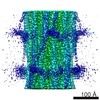
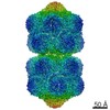
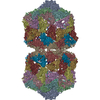
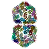
| タイトル | Structure of the helical Measles virus nucleocapsid | |||||||||
 マップデータ マップデータ | Reconstruction of trypsin digested Measles virus nucleocapsid | |||||||||
 試料 試料 |
| |||||||||
 キーワード キーワード | Measles virus Nucleocapsid / Transcription and Replication template | |||||||||
| 機能・相同性 |  機能・相同性情報 機能・相同性情報helical viral capsid / viral nucleocapsid / host cell cytoplasm / ribonucleoprotein complex / host cell nucleus / structural molecule activity / RNA binding 類似検索 - 分子機能 | |||||||||
| 生物種 |   Measles virus (ウイルス) Measles virus (ウイルス) | |||||||||
| 手法 | 単粒子再構成法 / クライオ電子顕微鏡法 / 解像度: 4.3 Å | |||||||||
 データ登録者 データ登録者 | Gutsche I / Desfosses A / Effantin G / Ling WL / Haupt M / Ruigrok RWH / Sachse C / Schoehn G | |||||||||
 引用 引用 | ジャーナル: Science / 年: 2015 タイトル: Structural virology. Near-atomic cryo-EM structure of the helical measles virus nucleocapsid. 著者: Irina Gutsche / Ambroise Desfosses / Grégory Effantin / Wai Li Ling / Melina Haupt / Rob W H Ruigrok / Carsten Sachse / Guy Schoehn /   要旨: Measles is a highly contagious human disease. We used cryo-electron microscopy and single particle-based helical image analysis to determine the structure of the helical nucleocapsid formed by the ...Measles is a highly contagious human disease. We used cryo-electron microscopy and single particle-based helical image analysis to determine the structure of the helical nucleocapsid formed by the folded domain of the measles virus nucleoprotein encapsidating an RNA at a resolution of 4.3 angstroms. The resulting pseudoatomic model of the measles virus nucleocapsid offers important insights into the mechanism of the helical polymerization of nucleocapsids of negative-strand RNA viruses, in particular via the exchange subdomains of the nucleoprotein. The structure reveals the mode of the nucleoprotein-RNA interaction and explains why each nucleoprotein of measles virus binds six nucleotides, whereas the respiratory syncytial virus nucleoprotein binds seven. It provides a rational basis for further analysis of measles virus replication and transcription, and reveals potential targets for drug design. | |||||||||
| 履歴 |
|
- 構造の表示
構造の表示
| ムービー |
ムービービューア |
|---|---|
| 構造ビューア | EMマップ: SurfViewMolmilJmol/JSmol |
| 添付画像 |

- ダウンロードとリンク
-EMDBアーカイブ
| マップデータ | emd_2867.map.gz | 30.3 MB | EMDBマップデータ形式 | |
|---|---|---|---|---|
| ヘッダ (付随情報) | emd-2867-v30.xmlemd-2867.xml | 10.3 KB 10.3 KB | 表示 表示 | EMDBヘッダ |
| 画像 |  EMD-2867-image.png EMD-2867-image.png | 426 KB | ||
| アーカイブディレクトリ |  http://ftp.pdbj.org/pub/emdb/structures/EMD-2867ftp://ftp.pdbj.org/pub/emdb/structures/EMD-2867 http://ftp.pdbj.org/pub/emdb/structures/EMD-2867ftp://ftp.pdbj.org/pub/emdb/structures/EMD-2867 | HTTPS FTP |
-関連構造データ


-リンク
| EMDBのページ | EMDB (EBI/PDBe) / EMDataResource |
|---|---|
| 「今月の分子」の関連する項目 |


-マップ
| ファイル | ダウンロード / ファイル: emd_2867.map.gz / 形式: CCP4 / 大きさ: 41.1 MB / タイプ: IMAGE STORED AS FLOATING POINT NUMBER (4 BYTES) | ||||||||||||||||||||||||||||||||||||||||||||||||||||||||||||
|---|---|---|---|---|---|---|---|---|---|---|---|---|---|---|---|---|---|---|---|---|---|---|---|---|---|---|---|---|---|---|---|---|---|---|---|---|---|---|---|---|---|---|---|---|---|---|---|---|---|---|---|---|---|---|---|---|---|---|---|---|---|
| 注釈 | Reconstruction of trypsin digested Measles virus nucleocapsid | ||||||||||||||||||||||||||||||||||||||||||||||||||||||||||||
| 投影像・断面図 | 画像のコントロール
画像は Spider により作成 これらの図は立方格子座標系で作成されたものです | ||||||||||||||||||||||||||||||||||||||||||||||||||||||||||||
| ボクセルのサイズ | X=Y=Z: 1.186 Å | ||||||||||||||||||||||||||||||||||||||||||||||||||||||||||||
| 密度 |
| ||||||||||||||||||||||||||||||||||||||||||||||||||||||||||||
| 対称性 | 空間群: 1 | ||||||||||||||||||||||||||||||||||||||||||||||||||||||||||||
| 詳細 | EMDB XML:
CCP4マップ ヘッダ情報:
| ||||||||||||||||||||||||||||||||||||||||||||||||||||||||||||
 Z (Sec.)
 Y (Row.)
 X (Col.)


-添付データ
- 試料の構成要素
-全体 : Recombinant truncated Measles Virus Nucleocapsid (Ncore-RNA helix)
| 全体 | 名称: Recombinant truncated Measles Virus Nucleocapsid (Ncore-RNA helix) |
|---|---|
| 要素 |
|
-超分子 #1000: Recombinant truncated Measles Virus Nucleocapsid (Ncore-RNA helix)
| 超分子 | 名称: Recombinant truncated Measles Virus Nucleocapsid (Ncore-RNA helix) タイプ: sample / ID: 1000 / 集合状態: Helical / Number unique components: 1 |
|---|
-分子 #1: Nucleoprotein
| 分子 | 名称: Nucleoprotein / タイプ: protein_or_peptide / ID: 1 / 詳細: Nucleoprotein was partially digested with trypsin / 集合状態: Helical / 組換発現: Yes |
|---|---|
| 由来(天然) | 生物種: Measles virus (ウイルス) / 株: Halle / 別称: Measles |
| 組換発現 | 生物種:   Spodoptera frugiperda (ツマジロクサヨトウ) Spodoptera frugiperda (ツマジロクサヨトウ)組換細胞: Sf21 |
-実験情報
-構造解析
| 手法 | クライオ電子顕微鏡法 |
|---|---|
 解析 解析 | 単粒子再構成法 |
| 試料の集合状態 | helical array |
-試料調製
| 緩衝液 | pH: 7.5 / 詳細: in 20 mM TriHCl pH 7.5, 150 mM NaCl |
|---|---|
| グリッド | 詳細: glow-discharged quantifoil grids 400 mesh 1.2/1.3 |
| 凍結 | 凍結剤: ETHANE / 装置: FEI VITROBOT MARK IV 手法: sample was applied to glow-discharged quantifoil grids 400 mesh 1.2/1.3, excess solution was blotted during 2 s with a Vitrobot Mark IV (FEI) and the grid frozen in liquid ethane |
- 電子顕微鏡法
| 顕微鏡 | FEI POLARA 300 |
|---|---|
| 詳細 | Special care was taken to perform a coma-free alignment of the microscope |
| 日付 | 2013年7月1日 |
| 撮影 | カテゴリ: FILM / フィルム・検出器のモデル: KODAK SO-163 FILM / デジタル化 - スキャナー: ZEISS SCAI / デジタル化 - サンプリング間隔: 7 µm |
| 電子線 | 加速電圧: 300 kV / 電子線源:  FIELD EMISSION GUN FIELD EMISSION GUN |
| 電子光学系 | 照射モード: FLOOD BEAM / 撮影モード: BRIGHT FIELD / 最大 デフォーカス(公称値): 0.0035 µm / 最小 デフォーカス(公称値): 0.0008 µm / 倍率(公称値): 59000 |
| 試料ステージ | 試料ホルダーモデル: GATAN LIQUID NITROGEN |
| 実験機器 |  モデル: Tecnai Polara / 画像提供: FEI Company |
-画像解析
| 詳細 | Final reconstruction obtained with SPRING |
|---|---|
| CTF補正 | 詳細: Each Particle |
| 最終 再構成 | 想定した対称性 - らせんパラメータ - Δz: 4.015 Å 想定した対称性 - らせんパラメータ - ΔΦ: 29.173 ° 想定した対称性 - らせんパラメータ - 軸対称性: C1 (非対称) アルゴリズム: OTHER / 解像度のタイプ: BY AUTHOR / 解像度: 4.3 Å / 解像度の算出法: OTHER / ソフトウェア - 名称: SPRING / 使用した粒子像数: 228165 |

