 Movie
Movie Controller

[English] 日本語
 Yorodumi
- PDB-6ek5: Near-atomic resolution structure of a plant geminivirus determine... -
+ Open data

- Basic information
| Entry | Database: PDB / ID: 6ek5 | |||||||||
|---|---|---|---|---|---|---|---|---|---|---|
| Title | Near-atomic resolution structure of a plant geminivirus determined by electron cryo-microscopy. | |||||||||
 Components Components | Capsid protein | |||||||||
 Keywords Keywords | VIRUS / African cassava mosaic virus / Geminivirus / ACMV | |||||||||
| Function / homology |  Function and homology information Function and homology informationT=1 icosahedral viral capsid / viral penetration into host nucleus / host cell / symbiont entry into host cell / host cell nucleus / structural molecule activity / DNA binding / metal ion binding Similarity search - Function | |||||||||
| Biological species |  African cassava mosaic virus African cassava mosaic virus | |||||||||
| Method | ELECTRON MICROSCOPY / single particle reconstruction / cryo EM / Resolution: 4.2 Å | |||||||||
 Authors Authors | Grimm, C. / Bottcher, B. / Hipp, K. / Jeske, H. | |||||||||
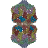
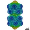
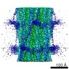
 Citation Citation | Journal: Structure / Year: 2017 Title: Near-Atomic Resolution Structure of a Plant Geminivirus Determined by Electron Cryomicroscopy. Authors: Katharina Hipp / Clemens Grimm / Holger Jeske / Bettina Böttcher /  Abstract: African cassava mosaic virus is a whitefly-transmitted geminivirus which forms unique twin particles of incomplete icosahedra that are joined at five-fold vertices, building an unusual waist. How its ...African cassava mosaic virus is a whitefly-transmitted geminivirus which forms unique twin particles of incomplete icosahedra that are joined at five-fold vertices, building an unusual waist. How its 22 capsomers interact within a half-capsid or across the waist is unknown thus far. Using electron cryo-microscopy and image processing, we determined the virion structure with a resolution of 4.2 Å and built an atomic model for its capsid protein. The inter-capsomer contacts mediated by the flexible N termini and loop regions differed within the half-capsids and at the waist, explaining partly the unusual twin structure. The tip of the pentameric capsomer is sealed by a plug formed by a turn region harboring the evolutionary conserved residue Y193. Basic amino acid residues inside the capsid form a positively charged pocket next to the five-fold axis of the capsomer suitable for binding DNA. Within this pocket, density most likely corresponding to DNA was resolved. | |||||||||
| History |
|
- Structure visualization
| Movie |
Movie viewer |
|---|---|
| Structure viewer | Molecule: MolmilJmol/JSmol |
- Downloads & links
-Download
| PDBx/mmCIF format | 6ek5.cif.gz | 3.5 MB | Display | PDBx/mmCIF format |
|---|---|---|---|---|
| PDB format | pdb6ek5.ent.gz | Display | PDB format | |
| PDBx/mmJSON format | 6ek5.json.gz | Tree view | PDBx/mmJSON format | |
| Others |  Other downloads Other downloads |
-Validation report
| Arichive directory | https://data.pdbj.org/pub/pdb/validation_reports/ek/6ek5ftp://data.pdbj.org/pub/pdb/validation_reports/ek/6ek5 | HTTPS FTP |
|---|
-Related structure data
| Related structure data |  3521 M: map data used to model this data |
|---|---|
| Similar structure data |

-Links
 PDBj
PDBj
- Assembly
| Deposited unit | 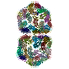
|
|---|---|
| 1 |
|
-Components
| #1: Protein | Mass: 24094.891 Da / Num. of mol.: 110 Source method: isolated from a genetically manipulated source Source: (gene. exp.) African cassava mosaic virus / Strain: isolate West Kenyan 844 / Gene: AR1, AV1 / Production host: African cassava mosaic virus / References: UniProt: P03561 |
|---|
-Experimental details
-Experiment
| Experiment | Method: ELECTRON MICROSCOPY |
|---|---|
| EM experiment | Aggregation state: PARTICLE / 3D reconstruction method: single particle reconstruction |
- Sample preparation
| Component | Name: African cassava mosaic virus - [West Kenya 844] / Type: VIRUS / Entity ID: all / Source: NATURAL |
|---|---|
| Molecular weight | Value: 3.3 MDa / Experimental value: NO |
| Source (natural) | Organism: African cassava mosaic virus - [West Kenya 844] |
| Details of virus | Empty: NO / Enveloped: NO / Isolate: STRAIN / Type: VIRION |
| Natural host | Organism: Manihot esculenta |
| Buffer solution | pH: 8 |
| Buffer component | Conc.: 0.1 M / Name: Sodium Borate / Formula: Na2B4O7 |
| Specimen | Embedding applied: NO / Shadowing applied: NO / Staining applied: NO / Vitrification applied: YES |
| Specimen support | Details: Quantifoil R1.2/1.3 + 2nm C. Glow discharged for 30-60 s with 25-28 MicroAmp (Quorum Tec Mini Sputter coater SC7620) and used within 1 hour Grid material: COPPER / Grid mesh size: 400 divisions/in. / Grid type: Quantifoil R1.2/1.3 |
| Vitrification | Instrument: FEI VITROBOT MARK IV / Cryogen name: ETHANE / Humidity: 100 % / Chamber temperature: 277 K Details: Samples (3 ?l) were applied to the glow discharged grids, incubated for 60 s on the grid, blotted and plunge frozen in liquid ethane using a Vitrobot IV (FEI, Eindhoven, The Netherlands) at ...Details: Samples (3 ?l) were applied to the glow discharged grids, incubated for 60 s on the grid, blotted and plunge frozen in liquid ethane using a Vitrobot IV (FEI, Eindhoven, The Netherlands) at 4?C with 100% humidity and blotting from both sides for 3 s with blot force 7 |
- Electron microscopy imaging
| Experimental equipment |  Model: Titan Krios / Image courtesy: FEI Company |
|---|---|
| Microscopy | Model: FEI TITAN KRIOS |
| Electron gun | Electron source:  FIELD EMISSION GUN / Accelerating voltage: 300 kV / Illumination mode: FLOOD BEAM FIELD EMISSION GUN / Accelerating voltage: 300 kV / Illumination mode: FLOOD BEAM |
| Electron lens | Mode: BRIGHT FIELD / Nominal magnification: 94000 X / Calibrated defocus min: 780 nm / Calibrated defocus max: 5600 nm / Cs: 0.01 mm / Alignment procedure: COMA FREE |
| Specimen holder | Cryogen: NITROGEN / Specimen holder model: FEI TITAN KRIOS AUTOGRID HOLDER |
| Image recording | Electron dose: 25 e/Å2 / Detector mode: INTEGRATING / Film or detector model: FEI FALCON II (4k x 4k) / Num. of grids imaged: 1 / Num. of real images: 934 Details: data acquisition with a cs-corrected FEI Titan Krios on a Falcon II direct detector at 300 kV. Data was acquired at a primary magnification of 94,000 (calibrated pixel size of 1.57 A) and ...Details: data acquisition with a cs-corrected FEI Titan Krios on a Falcon II direct detector at 300 kV. Data was acquired at a primary magnification of 94,000 (calibrated pixel size of 1.57 A) and with a total dose of 25 e/A2 in 17 frames. In total 1,108 movies were recorded of which 934 movies were used for further processing. The movie frames were averaged after motion correction and dose weightin |
| EM imaging optics | Spherical aberration corrector: Krios - cs-corrector |
| Image scans | Width: 4096 / Height: 4096 / Movie frames/image: 17 / Used frames/image: 1-17 |
- Processing
| EM software |
| ||||||||||||||||||||||||||||||||||||
|---|---|---|---|---|---|---|---|---|---|---|---|---|---|---|---|---|---|---|---|---|---|---|---|---|---|---|---|---|---|---|---|---|---|---|---|---|---|
| Image processing | Details: Images were motion correted and weighted for dose damage; the CTF was determined with CTFFIND 3 | ||||||||||||||||||||||||||||||||||||
| CTF correction | Type: PHASE FLIPPING AND AMPLITUDE CORRECTION | ||||||||||||||||||||||||||||||||||||
| Particle selection | Num. of particles selected: 141141 Details: For the initial analysis, ca. 2,000 particle images were selected manually with e2boxer and extracted and normalized with relion 1.4 followed by 2D-classification and ctf-correction. Three ...Details: For the initial analysis, ca. 2,000 particle images were selected manually with e2boxer and extracted and normalized with relion 1.4 followed by 2D-classification and ctf-correction. Three characteristic class averages that resolved the two parts of the twin particle (side views and intermediate views) were selected as references for template-dependent automatic particle picking in Relion. Particle images were extracted at the determined coordinates with a box size of 300 x 300 Px and normalized for their grey value distribution followed by 2D-classification. Some of the 2D-classes showed disconnected twin particles or single capsids. These classes probably represented either adjacent capsids from different twin particles or incorrectly centered particles and were excluded from the subsequent analysis. 141141 particles is the number of automatically selected particles; 69685 were retained for he subsequent processing | ||||||||||||||||||||||||||||||||||||
| Symmetry | Point symmetry: D5 (2x5 fold dihedral) | ||||||||||||||||||||||||||||||||||||
| 3D reconstruction | Resolution: 4.2 Å / Resolution method: FSC 0.143 CUT-OFF / Num. of particles: 24451 / Algorithm: FOURIER SPACE / Num. of class averages: 1 / Symmetry type: POINT | ||||||||||||||||||||||||||||||||||||
| Atomic model building | B value: 139 / Protocol: FLEXIBLE FIT / Space: REAL | ||||||||||||||||||||||||||||||||||||
| Refinement | Highest resolution: 4.2 Å |
