 Movie
Movie Controller


+ Open data

- Basic information
| Entry | Database: PDB / ID: 6f2s | ||||||||||||||||||
|---|---|---|---|---|---|---|---|---|---|---|---|---|---|---|---|---|---|---|---|
| Title | CryoEM structure of Ageratum Yellow Vein virus (AYVV) | ||||||||||||||||||
 Components Components |
| ||||||||||||||||||
 Keywords Keywords | VIRUS / AYVV / geminivirus / ssDNA / gemini | ||||||||||||||||||
| Function / homology |  Function and homology information Function and homology informationT=1 icosahedral viral capsid / viral penetration into host nucleus / host cell / symbiont entry into host cell / host cell nucleus / structural molecule activity / DNA binding / zinc ion binding Similarity search - Function | ||||||||||||||||||
| Biological species |  Ageratum yellow vein virus Ageratum yellow vein virus | ||||||||||||||||||
| Method | ELECTRON MICROSCOPY / single particle reconstruction / cryo EM / Resolution: 3.3 Å | ||||||||||||||||||
 Authors Authors | Hesketh, E.L. / Saunders, K. / Fisher, C. / Potze, J. / Stanley, J. / Lomonossoff, G.P. / Ranson, N.A. | ||||||||||||||||||
| Funding support |  United Kingdom, 5items United Kingdom, 5items
| ||||||||||||||||||
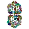
 Citation Citation | Journal: Nat Commun / Year: 2018 Title: The 3.3 Å structure of a plant geminivirus using cryo-EM. Authors: Emma L Hesketh / Keith Saunders / Chloe Fisher / Joran Potze / John Stanley / George P Lomonossoff / Neil A Ranson / Abstract: Geminiviruses are major plant pathogens that threaten food security globally. They have a unique architecture built from two incomplete icosahedral particles, fused to form a geminate capsid. ...Geminiviruses are major plant pathogens that threaten food security globally. They have a unique architecture built from two incomplete icosahedral particles, fused to form a geminate capsid. However, despite their importance to agricultural economies and fundamental biological interest, the details of how this is realized in 3D remain unknown. Here we report the structure of Ageratum yellow vein virus at 3.3 Å resolution, using single-particle cryo-electron microscopy, together with an atomic model that shows that the N-terminus of the single capsid protein (CP) adopts three different conformations essential for building the interface between geminate halves. Our map also contains density for ~7 bases of single-stranded DNA bound to each CP, and we show that the interactions between the genome and CPs are different at the interface than in the rest of the capsid. With additional mutagenesis data, this suggests a central role for DNA binding-induced conformational change in directing the assembly of geminate capsids. | ||||||||||||||||||
| History |
|
- Structure visualization
| Movie |
Movie viewer |
|---|---|
| Structure viewer | Molecule: MolmilJmol/JSmol |
- Downloads & links
-Download
| PDBx/mmCIF format | 6f2s.cif.gz | 469 KB | Display | PDBx/mmCIF format |
|---|---|---|---|---|
| PDB format | pdb6f2s.ent.gz | 384.8 KB | Display | PDB format |
| PDBx/mmJSON format | 6f2s.json.gz | Tree view | PDBx/mmJSON format | |
| Others |  Other downloads Other downloads |
-Validation report
| Arichive directory | https://data.pdbj.org/pub/pdb/validation_reports/f2/6f2sftp://data.pdbj.org/pub/pdb/validation_reports/f2/6f2s | HTTPS FTP |
|---|
-Related structure data
| Related structure data | 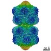 4174MC M: map data used to model this data C: citing same article ( |
|---|---|
| Similar structure data |

-Links
 PDBj
PDBj
- Assembly
| Deposited unit | 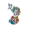
|
|---|---|
| 1 | x 10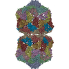
|
| 2 |
|
| 3 | 
|
| Symmetry | Point symmetry: (Schoenflies symbol: D5 (2x5 fold dihedral)) |
-Components
| #1: Protein | Mass: 22500.703 Da / Num. of mol.: 9 Source method: isolated from a genetically manipulated source Source: (gene. exp.) Ageratum yellow vein virus / Gene: V1 / Production host:  #2: DNA chain | Mass: 2051.390 Da / Num. of mol.: 10 Source method: isolated from a genetically manipulated source Source: (gene. exp.) Ageratum yellow vein virus / Production host: #3: Protein | | Mass: 23644.094 Da / Num. of mol.: 1 Source method: isolated from a genetically manipulated source Source: (gene. exp.) Ageratum yellow vein virus / Gene: V1 / Production host: #4: Protein | | Mass: 25664.473 Da / Num. of mol.: 1 Source method: isolated from a genetically manipulated source Source: (gene. exp.) Ageratum yellow vein virus / Gene: V1 / Production host: #5: DNA chain | | Mass: 1762.208 Da / Num. of mol.: 1 Source method: isolated from a genetically manipulated source Source: (gene. exp.) Ageratum yellow vein virus / Production host: |
|---|
-Experimental details
-Experiment
| Experiment | Method: ELECTRON MICROSCOPY |
|---|---|
| EM experiment | Aggregation state: PARTICLE / 3D reconstruction method: single particle reconstruction |
- Sample preparation
| Component | Name: Ageratum yellow vein virus / Type: VIRUS / Entity ID: all / Source: NATURAL |
|---|---|
| Molecular weight | Value: 0.251 MDa |
| Source (natural) | Organism: Ageratum yellow vein virus |
| Source (recombinant) | Organism: |
| Details of virus | Empty: NO / Enveloped: NO / Isolate: SPECIES / Type: VIRION |
| Natural host | Organism: Ageratum |
| Buffer solution | pH: 7 |
| Buffer component | Conc.: 100 mM / Name: Sodium phosphate / Formula: NaPo4 |
| Specimen | Embedding applied: NO / Shadowing applied: NO / Staining applied: NO / Vitrification applied: YES |
| Specimen support | Details: PELCO easiglow / Grid material: COPPER / Grid mesh size: 400 divisions/in. |
| Vitrification | Instrument: LEICA EM GP / Cryogen name: ETHANE / Humidity: 90 % / Chamber temperature: 277 K |
- Electron microscopy imaging
| Experimental equipment |  Model: Titan Krios / Image courtesy: FEI Company |
|---|---|
| Microscopy | Model: FEI TITAN KRIOS |
| Electron gun | Electron source:  FIELD EMISSION GUN / Accelerating voltage: 300 kV / Illumination mode: SPOT SCAN FIELD EMISSION GUN / Accelerating voltage: 300 kV / Illumination mode: SPOT SCAN |
| Electron lens | Mode: BRIGHT FIELD / Nominal magnification: 75000 X / Nominal defocus max: 2700 nm / Nominal defocus min: 800 nm / Calibrated defocus min: 300 nm / Calibrated defocus max: 5000 nm / Cs: 2.7 mm / C2 aperture diameter: 70 µm / Alignment procedure: COMA FREE |
| Specimen holder | Cryogen: NITROGEN / Specimen holder model: FEI TITAN KRIOS AUTOGRID HOLDER / Temperature (max): 78 K / Temperature (min): 78 K |
| Image recording | Average exposure time: 2 sec. / Electron dose: 110 e/Å2 / Detector mode: INTEGRATING / Film or detector model: FEI FALCON III (4k x 4k) / Num. of grids imaged: 1 / Num. of real images: 12028 |
- Processing
| Software | Name: REFMAC / Version: 5.8.0107 / Classification: refinement | ||||||||||||||||||||||||||||||||||||||||||||||||||||||||||||||||||||||||||||||||||||||||||||||||||||||||||
|---|---|---|---|---|---|---|---|---|---|---|---|---|---|---|---|---|---|---|---|---|---|---|---|---|---|---|---|---|---|---|---|---|---|---|---|---|---|---|---|---|---|---|---|---|---|---|---|---|---|---|---|---|---|---|---|---|---|---|---|---|---|---|---|---|---|---|---|---|---|---|---|---|---|---|---|---|---|---|---|---|---|---|---|---|---|---|---|---|---|---|---|---|---|---|---|---|---|---|---|---|---|---|---|---|---|---|---|
| EM software |
| ||||||||||||||||||||||||||||||||||||||||||||||||||||||||||||||||||||||||||||||||||||||||||||||||||||||||||
| CTF correction | Type: PHASE FLIPPING AND AMPLITUDE CORRECTION | ||||||||||||||||||||||||||||||||||||||||||||||||||||||||||||||||||||||||||||||||||||||||||||||||||||||||||
| Particle selection | Num. of particles selected: 116240 | ||||||||||||||||||||||||||||||||||||||||||||||||||||||||||||||||||||||||||||||||||||||||||||||||||||||||||
| Symmetry | Point symmetry: D5 (2x5 fold dihedral) | ||||||||||||||||||||||||||||||||||||||||||||||||||||||||||||||||||||||||||||||||||||||||||||||||||||||||||
| 3D reconstruction | Resolution: 3.3 Å / Resolution method: FSC 0.143 CUT-OFF / Num. of particles: 64932 / Symmetry type: POINT | ||||||||||||||||||||||||||||||||||||||||||||||||||||||||||||||||||||||||||||||||||||||||||||||||||||||||||
| Atomic model building | Protocol: RIGID BODY FIT | ||||||||||||||||||||||||||||||||||||||||||||||||||||||||||||||||||||||||||||||||||||||||||||||||||||||||||
| Refinement | Resolution: 3.3→477.16 Å / Cor.coef. Fo:Fc: 0.8 / SU B: 20.039 / SU ML: 0.273 / ESU R: 0.061 Stereochemistry target values: MAXIMUM LIKELIHOOD WITH PHASES Details: HYDROGENS HAVE BEEN ADDED IN THE RIDING POSITIONS
| ||||||||||||||||||||||||||||||||||||||||||||||||||||||||||||||||||||||||||||||||||||||||||||||||||||||||||
| Solvent computation | Ion probe radii: 0.8 Å / Shrinkage radii: 0.8 Å / VDW probe radii: 1.2 Å / Solvent model: MASK | ||||||||||||||||||||||||||||||||||||||||||||||||||||||||||||||||||||||||||||||||||||||||||||||||||||||||||
| Displacement parameters | Biso mean: 97.466 Å2
| ||||||||||||||||||||||||||||||||||||||||||||||||||||||||||||||||||||||||||||||||||||||||||||||||||||||||||
| Refinement step | Cycle: 1 / Total: 19170 | ||||||||||||||||||||||||||||||||||||||||||||||||||||||||||||||||||||||||||||||||||||||||||||||||||||||||||
| Refine LS restraints |
|