 Movie
Movie Controller


+ Open data

- Basic information
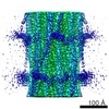
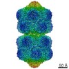
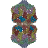
| Entry | Database: EMDB / ID: EMD-2867 | |||||||||
|---|---|---|---|---|---|---|---|---|---|---|
| Title | Structure of the helical Measles virus nucleocapsid | |||||||||
 Map data Map data | Reconstruction of trypsin digested Measles virus nucleocapsid | |||||||||
 Sample Sample |
| |||||||||
 Keywords Keywords | Measles virus Nucleocapsid / Transcription and Replication template | |||||||||
| Function / homology |  Function and homology information Function and homology informationhelical viral capsid / viral nucleocapsid / host cell cytoplasm / ribonucleoprotein complex / host cell nucleus / structural molecule activity / RNA binding Similarity search - Function | |||||||||
| Biological species |   Measles virus Measles virus | |||||||||
| Method | single particle reconstruction / cryo EM / Resolution: 4.3 Å | |||||||||
 Authors Authors | Gutsche I / Desfosses A / Effantin G / Ling WL / Haupt M / Ruigrok RWH / Sachse C / Schoehn G | |||||||||
 Citation Citation | Journal: Science / Year: 2015 Title: Structural virology. Near-atomic cryo-EM structure of the helical measles virus nucleocapsid. Authors: Irina Gutsche / Ambroise Desfosses / Grégory Effantin / Wai Li Ling / Melina Haupt / Rob W H Ruigrok / Carsten Sachse / Guy Schoehn /   Abstract: Measles is a highly contagious human disease. We used cryo-electron microscopy and single particle-based helical image analysis to determine the structure of the helical nucleocapsid formed by the ...Measles is a highly contagious human disease. We used cryo-electron microscopy and single particle-based helical image analysis to determine the structure of the helical nucleocapsid formed by the folded domain of the measles virus nucleoprotein encapsidating an RNA at a resolution of 4.3 angstroms. The resulting pseudoatomic model of the measles virus nucleocapsid offers important insights into the mechanism of the helical polymerization of nucleocapsids of negative-strand RNA viruses, in particular via the exchange subdomains of the nucleoprotein. The structure reveals the mode of the nucleoprotein-RNA interaction and explains why each nucleoprotein of measles virus binds six nucleotides, whereas the respiratory syncytial virus nucleoprotein binds seven. It provides a rational basis for further analysis of measles virus replication and transcription, and reveals potential targets for drug design. | |||||||||
| History |
|
- Structure visualization
| Movie |
Movie viewer |
|---|---|
| Structure viewer | EM map: SurfViewMolmilJmol/JSmol |
| Supplemental images |

- Downloads & links
-EMDB archive
| Map data | emd_2867.map.gz | 30.3 MB | EMDB map data format | |
|---|---|---|---|---|
| Header (meta data) | emd-2867-v30.xmlemd-2867.xml | 10.3 KB 10.3 KB | Display Display | EMDB header |
| Images |  EMD-2867-image.png EMD-2867-image.png | 426 KB | ||
| Archive directory |  http://ftp.pdbj.org/pub/emdb/structures/EMD-2867ftp://ftp.pdbj.org/pub/emdb/structures/EMD-2867 http://ftp.pdbj.org/pub/emdb/structures/EMD-2867ftp://ftp.pdbj.org/pub/emdb/structures/EMD-2867 | HTTPS FTP |
-Related structure data
| Related structure data |  4uftMC M: atomic model generated by this map C: citing same article ( |
|---|---|
| Similar structure data |


-Links
| EMDB pages | EMDB (EBI/PDBe) / EMDataResource |
|---|---|
| Related items in Molecule of the Month |


-Map
| File | Download / File: emd_2867.map.gz / Format: CCP4 / Size: 41.1 MB / Type: IMAGE STORED AS FLOATING POINT NUMBER (4 BYTES) | ||||||||||||||||||||||||||||||||||||||||||||||||||||||||||||
|---|---|---|---|---|---|---|---|---|---|---|---|---|---|---|---|---|---|---|---|---|---|---|---|---|---|---|---|---|---|---|---|---|---|---|---|---|---|---|---|---|---|---|---|---|---|---|---|---|---|---|---|---|---|---|---|---|---|---|---|---|---|
| Annotation | Reconstruction of trypsin digested Measles virus nucleocapsid | ||||||||||||||||||||||||||||||||||||||||||||||||||||||||||||
| Projections & slices | Image control
Images are generated by Spider. generated in cubic-lattice coordinate | ||||||||||||||||||||||||||||||||||||||||||||||||||||||||||||
| Voxel size | X=Y=Z: 1.186 Å | ||||||||||||||||||||||||||||||||||||||||||||||||||||||||||||
| Density |
| ||||||||||||||||||||||||||||||||||||||||||||||||||||||||||||
| Symmetry | Space group: 1 | ||||||||||||||||||||||||||||||||||||||||||||||||||||||||||||
| Details | EMDB XML:
CCP4 map header:
| ||||||||||||||||||||||||||||||||||||||||||||||||||||||||||||
 Z (Sec.)
 Y (Row.)
 X (Col.)


-Supplemental data
- Sample components
-Entire : Recombinant truncated Measles Virus Nucleocapsid (Ncore-RNA helix)
| Entire | Name: Recombinant truncated Measles Virus Nucleocapsid (Ncore-RNA helix) |
|---|---|
| Components |
|
-Supramolecule #1000: Recombinant truncated Measles Virus Nucleocapsid (Ncore-RNA helix)
| Supramolecule | Name: Recombinant truncated Measles Virus Nucleocapsid (Ncore-RNA helix) type: sample / ID: 1000 / Oligomeric state: Helical / Number unique components: 1 |
|---|
-Macromolecule #1: Nucleoprotein
| Macromolecule | Name: Nucleoprotein / type: protein_or_peptide / ID: 1 / Details: Nucleoprotein was partially digested with trypsin / Oligomeric state: Helical / Recombinant expression: Yes |
|---|---|
| Source (natural) | Organism: Measles virus / Strain: Halle / synonym: Measles |
| Recombinant expression | Organism:   Spodoptera frugiperda (fall armyworm) / Recombinant cell: Sf21 Spodoptera frugiperda (fall armyworm) / Recombinant cell: Sf21 |
-Experimental details
-Structure determination
| Method | cryo EM |
|---|---|
 Processing Processing | single particle reconstruction |
| Aggregation state | helical array |
-Sample preparation
| Buffer | pH: 7.5 / Details: in 20 mM TriHCl pH 7.5, 150 mM NaCl |
|---|---|
| Grid | Details: glow-discharged quantifoil grids 400 mesh 1.2/1.3 |
| Vitrification | Cryogen name: ETHANE / Instrument: FEI VITROBOT MARK IV Method: sample was applied to glow-discharged quantifoil grids 400 mesh 1.2/1.3, excess solution was blotted during 2 s with a Vitrobot Mark IV (FEI) and the grid frozen in liquid ethane |
- Electron microscopy
| Microscope | FEI POLARA 300 |
|---|---|
| Details | Special care was taken to perform a coma-free alignment of the microscope |
| Date | Jul 1, 2013 |
| Image recording | Category: FILM / Film or detector model: KODAK SO-163 FILM / Digitization - Scanner: ZEISS SCAI / Digitization - Sampling interval: 7 µm |
| Electron beam | Acceleration voltage: 300 kV / Electron source:  FIELD EMISSION GUN FIELD EMISSION GUN |
| Electron optics | Illumination mode: FLOOD BEAM / Imaging mode: BRIGHT FIELD / Nominal defocus max: 0.0035 µm / Nominal defocus min: 0.0008 µm / Nominal magnification: 59000 |
| Sample stage | Specimen holder model: GATAN LIQUID NITROGEN |
| Experimental equipment |  Model: Tecnai Polara / Image courtesy: FEI Company |
-Image processing
| Details | Final reconstruction obtained with SPRING |
|---|---|
| CTF correction | Details: Each Particle |
| Final reconstruction | Applied symmetry - Helical parameters - Δz: 4.015 Å Applied symmetry - Helical parameters - Δ&Phi: 29.173 ° Applied symmetry - Helical parameters - Axial symmetry: C1 (asymmetric) Algorithm: OTHER / Resolution.type: BY AUTHOR / Resolution: 4.3 Å / Resolution method: OTHER / Software - Name: SPRING / Number images used: 228165 |

