[English] 日本語
Yorodumi
- EMDB-2705: A structural model of the active ribosome-bound membrane protein ... -

+
Open data


ID or keywords:

Loading...

-
Basic information

Entry
Database: EMDB / ID: EMD-2705
TitleA structural model of the active ribosome-bound membrane protein insertase YidC
Map dataCryo-em reconstruction of YidC bound a ribosome nascent chain complex
Sample
  • Sample: Monomeric YidC bound to ribosome nascent chain complex(F0c)
  • Complex: Ribosome nascent chain complexRibosome-nascent chain complex
  • Protein or peptide: YidC
Keywordsribosome / YidC / protein translocation / bioinformatics / MD simulation / membrane
Function / homology
Function and homology information


protein insertion into membrane from inner side / membrane insertase activity / cell envelope Sec protein transport complex / protein transport by the Sec complex / proton-transporting ATP synthase complex / proton motive force-driven plasma membrane ATP synthesis / protein insertion into membrane / proton-transporting ATP synthase complex, coupling factor F(o) / proton motive force-driven ATP synthesis / proton-transporting ATPase activity, rotational mechanism ...protein insertion into membrane from inner side / membrane insertase activity / cell envelope Sec protein transport complex / protein transport by the Sec complex / proton-transporting ATP synthase complex / proton motive force-driven plasma membrane ATP synthesis / protein insertion into membrane / proton-transporting ATP synthase complex, coupling factor F(o) / proton motive force-driven ATP synthesis / proton-transporting ATPase activity, rotational mechanism / proton-transporting ATP synthase activity, rotational mechanism / protein folding / protein-containing complex assembly / lipid binding / membrane / plasma membrane
Similarity search - Function
Membrane insertase YidC, N-terminal / YidC, periplasmic domain superfamily / YidC periplasmic domain / : / Membrane insertase YidC / Membrane insertase YidC/Oxa1, C-terminal / 60Kd inner membrane protein / Membrane insertase YidC/ALB3/OXA1/COX18 / ATP synthase, F0 complex, subunit C, bacterial/chloroplast / ATP synthase, F0 complex, subunit C ...Membrane insertase YidC, N-terminal / YidC, periplasmic domain superfamily / YidC periplasmic domain / : / Membrane insertase YidC / Membrane insertase YidC/Oxa1, C-terminal / 60Kd inner membrane protein / Membrane insertase YidC/ALB3/OXA1/COX18 / ATP synthase, F0 complex, subunit C, bacterial/chloroplast / ATP synthase, F0 complex, subunit C / F1F0 ATP synthase subunit C superfamily / ATP synthase, F0 complex, subunit C, DCCD-binding site / ATP synthase c subunit signature. / V-ATPase proteolipid subunit C-like domain / F/V-ATP synthase subunit C superfamily / ATP synthase subunit C
Similarity search - Domain/homology
Membrane protein insertase YidC / ATP synthase subunit c
Similarity search - Component
Biological speciesEscherichia coli (E. coli)
Methodsingle particle reconstruction / cryo EM / Resolution: 8.0 Å
AuthorsWickles S / Singharoy A / Andreani J / Seemayer S / Bischoff L / Berninghausen O / Soeding J / Schulten K / van der Sluis EO / Beckmann R
Citation
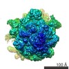
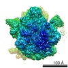
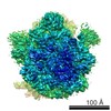
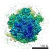
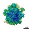
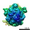
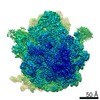
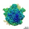
Journal: Elife / Year: 2014
Title: A structural model of the active ribosome-bound membrane protein insertase YidC.
Authors: Stephan Wickles / Abhishek Singharoy / Jessica Andreani / Stefan Seemayer / Lukas Bischoff / Otto Berninghausen / Johannes Soeding / Klaus Schulten / Eli O van der Sluis / Roland Beckmann /
Abstract: The integration of most membrane proteins into the cytoplasmic membrane of bacteria occurs co-translationally. The universally conserved YidC protein mediates this process either individually as a ...The integration of most membrane proteins into the cytoplasmic membrane of bacteria occurs co-translationally. The universally conserved YidC protein mediates this process either individually as a membrane protein insertase, or in concert with the SecY complex. Here, we present a structural model of YidC based on evolutionary co-variation analysis, lipid-versus-protein-exposure and molecular dynamics simulations. The model suggests a distinctive arrangement of the conserved five transmembrane domains and a helical hairpin between transmembrane segment 2 (TM2) and TM3 on the cytoplasmic membrane surface. The model was used for docking into a cryo-electron microscopy reconstruction of a translating YidC-ribosome complex carrying the YidC substrate FOc. This structure reveals how a single copy of YidC interacts with the ribosome at the ribosomal tunnel exit and identifies a site for membrane protein insertion at the YidC protein-lipid interface. Together, these data suggest a mechanism for the co-translational mode of YidC-mediated membrane protein insertion.
History
DepositionJul 10, 2014-
Header (metadata) releaseJul 23, 2014-
Map releaseJul 23, 2014-
UpdateAug 27, 2014-
Current statusAug 27, 2014Processing site: PDBe / Status: Released

-
Structure visualization
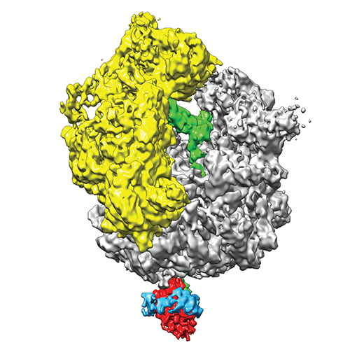
Movie
  • Surface view with section colored by density value
  • Surface level: 0.35
  • Imaged by UCSF Chimera
  • Download
  • Surface view colored by height
  • Surface level: 0.35
  • Imaged by UCSF Chimera
  • Download
  • Surface view with fitted model
  • Atomic models: PDB-4utq
  • Surface level: 0.35
  • Imaged by UCSF Chimera
  • Download
  • Surface view with fitted model
  • Atomic models: PDB-4utq
  • Surface level: 0.35
  • Imaged by UCSF Chimera
  • Download
  • Simplified surface model + fitted atomic model
  • Atomic modelsPDB-4utq
  • Imaged by Jmol
  • Download
Movie viewer
Structure viewerEM map:
SurfViewMolmilJmol/JSmol
Supplemental images

Downloads & links

-
Map

FileDownload / File: emd_2705.map.gz / Format: CCP4 / Size: 185.7 MB / Type: IMAGE STORED AS FLOATING POINT NUMBER (4 BYTES)
AnnotationCryo-em reconstruction of YidC bound a ribosome nascent chain complex
Voxel sizeX=Y=Z: 1.035 Å
Density
Contour LevelBy EMDB: 0.25 / Movie #1: 0.35
Minimum - Maximum-0.53752744 - 1.26601863
Average (Standard dev.)0.00298494 (±0.13338423)
SymmetrySpace group: 1
Details

EMDB XML:

Map geometry
Axis orderXYZ
Origin-184-184-183
Dimensions368368368
Spacing368368368
CellA=B=C: 380.87997 Å
α=β=γ: 90.0 °

CCP4 map header:

modeImage stored as Reals
Å/pix. X/Y/Z1.0351.0351.035
M x/y/z368368368
origin x/y/z0.0000.0000.000
length x/y/z380.880380.880380.880
α/β/γ90.00090.00090.000
start NX/NY/NZ-180-180-179
NX/NY/NZ360360360
MAP C/R/S123
start NC/NR/NS-184-184-183
NC/NR/NS368368368
D min/max/mean-0.5381.2660.003

-
Supplemental data

-
Sample components

-
Entire : Monomeric YidC bound to ribosome nascent chain complex(F0c)

EntireName: Monomeric YidC bound to ribosome nascent chain complex(F0c)
Components
  • Sample: Monomeric YidC bound to ribosome nascent chain complex(F0c)
  • Complex: Ribosome nascent chain complexRibosome-nascent chain complex
  • Protein or peptide: YidC

-
Supramolecule #1000: Monomeric YidC bound to ribosome nascent chain complex(F0c)

SupramoleculeName: Monomeric YidC bound to ribosome nascent chain complex(F0c)
type: sample / ID: 1000 / Number unique components: 2

-
Supramolecule #1: Ribosome nascent chain complex

SupramoleculeName: Ribosome nascent chain complex / type: complex / ID: 1 / Recombinant expression: No / Ribosome-details: ribosome-prokaryote: ALL
Source (natural)Organism: Escherichia coli (E. coli)

-
Macromolecule #1: YidC

MacromoleculeName: YidC / type: protein_or_peptide / ID: 1 / Recombinant expression: No
Source (natural)Organism: Escherichia coli (E. coli)

-
Experimental details

-
Structure determination

Methodcryo EM
Processingsingle particle reconstruction
Aggregation stateparticle

-
Sample preparation

VitrificationCryogen name: ETHANE / Instrument: FEI VITROBOT MARK IV

-
Electron microscopy

MicroscopeFEI TITAN KRIOS
Electron beamAcceleration voltage: 200 kV / Electron source: FIELD EMISSION GUN
Electron opticsIllumination mode: SPOT SCAN / Imaging mode: BRIGHT FIELDBright-field microscopy / Nominal defocus max: 3.5 µm / Nominal defocus min: 1.3 µm
Sample stageSpecimen holder model: FEI TITAN KRIOS AUTOGRID HOLDER
DateJun 1, 2013
Image recordingCategory: CCD / Film or detector model: TVIPS TEMCAM-F416 (4k x 4k) / Number real images: 9999 / Average electron dose: 20 e/Å2
Experimental equipment
Model: Titan Krios / Image courtesy: FEI Company

-
Image processing

CTF correctionDetails: Defocus group
Final reconstructionApplied symmetry - Point group: C1 (asymmetric) / Resolution.type: BY AUTHOR / Resolution: 8.0 Å / Resolution method: OTHER / Software - Name: Spider / Number images used: 58960

+
About Yorodumi

-
News

-
Feb 9, 2022. New format data for meta-information of EMDB entries

New format data for meta-information of EMDB entries

  • Version 3 of the EMDB header file is now the official format.
  • The previous official version 1.9 will be removed from the archive.

Related info.:EMDB header

External links:wwPDB to switch to version 3 of the EMDB data model

-
Aug 12, 2020. Covid-19 info

Covid-19 info

URL: https://pdbj.org/emnavi/covid19.php

New page: Covid-19 featured information page in EM Navigator.

Related info.:Covid-19 info / Mar 5, 2020. Novel coronavirus structure data

+
Mar 5, 2020. Novel coronavirus structure data

Novel coronavirus structure data

Related info.:Yorodumi Speices / Aug 12, 2020. Covid-19 info

External links:COVID-19 featured content - PDBj / Molecule of the Month (242):Coronavirus Proteases

+
Jan 31, 2019. EMDB accession codes are about to change! (news from PDBe EMDB page)

EMDB accession codes are about to change! (news from PDBe EMDB page)

  • The allocation of 4 digits for EMDB accession codes will soon come to an end. Whilst these codes will remain in use, new EMDB accession codes will include an additional digit and will expand incrementally as the available range of codes is exhausted. The current 4-digit format prefixed with “EMD-” (i.e. EMD-XXXX) will advance to a 5-digit format (i.e. EMD-XXXXX), and so on. It is currently estimated that the 4-digit codes will be depleted around Spring 2019, at which point the 5-digit format will come into force.
  • The EM Navigator/Yorodumi systems omit the EMD- prefix.

Related info.:Q: What is EMD? / ID/Accession-code notation in Yorodumi/EM Navigator

External links:EMDB Accession Codes are Changing Soon! / Contact to PDBj

+
Jul 12, 2017. Major update of PDB

Major update of PDB

  • wwPDB released updated PDB data conforming to the new PDBx/mmCIF dictionary.
  • This is a major update changing the version number from 4 to 5, and with Remediation, in which all the entries are updated.
  • In this update, many items about electron microscopy experimental information are reorganized (e.g. em_software).
  • Now, EM Navigator and Yorodumi are based on the updated data.

External links:wwPDB Remediation / Enriched Model Files Conforming to OneDep Data Standards Now Available in the PDB FTP Archive

-
Yorodumi

Thousand views of thousand structures

  • Yorodumi is a browser for structure data from EMDB, PDB, SASBDB, etc.
  • This page is also the successor to EM Navigator detail page, and also detail information page/front-end page for Omokage search.
  • The word "yorodu" (or yorozu) is an old Japanese word meaning "ten thousand". "mi" (miru) is to see.

Related info.:EMDB / PDB / SASBDB / Comparison of 3 databanks / Yorodumi Search / Aug 31, 2016. New EM Navigator & Yorodumi / Yorodumi Papers / Jmol/JSmol / Function and homology information / Changes in new EM Navigator and Yorodumi

Read more